SESSIONE E-POSTER 1 - ARITMOLOGIA CLINICA E INTERVENTISTICA - AIAC
←
→
Trascrizione del contenuto della pagina
Se il tuo browser non visualizza correttamente la pagina, ti preghiamo di leggere il contenuto della pagina quaggiù
CONGRESSO
       NAZIONALE

     AIAC                                                                                                                              Bologna
                                                                                                                                       10-12 Marzo 2016
GIOVEDI’ 10 MARZO 2016

13.30 - 14.00
AREA POSTER
SESSIONE E-POSTER 1 - ARITMOLOGIA CLINICA E INTERVENTISTICA

01
VENTRICULAR ARRHYTHMIAS IN SUSPECTED UNDERLYING CARDIOMYOPATHY: ROLE OF ENDOMIOCARDIAL
BIOPSY GUIDED BY BIPOLAR-UNIPOLAR VOLTAGE MAPPING
G. Vettor 1, A. Dello Russo 1, M. Casella 1, G.M. Fassini 1, S. Riva 1, M. Moltrasio 1, F. Tundo 1, B. Majocchi 1, M. Zucchetti 1, V. Marino 1, S. Conti 1,
F. Pizzamiglio 1, M.A. Dessanai 1, E. Russo 1, C. Carbucicchio 1, C. Basso 2, G. Thiene 2, C. Tondo1
1
  Cardiac Arrhythmia Center Centro Cardiologico Monzino, Milano, ITALY
2
  Cardiovascular Pathology Università di Padova, Padova, ITALY

Introduction: Discriminate between idiopathic ventricular arrhythmias and arrhythmia sign of an underlying cardiomyopathy is a challenge in the
clinical practice. We evaluated the role of endomiocardial biopsy (EMB) guided by electroanatomical mapping in the management of patients with
ventricular arrhythmias in terms of diagnosis of underlying cardiomyopathy, risk stratification and therapeutic strategy.

Methods: From January 2010 to October 2015, 60 consecutive patients with frequent or sporadic ventricular arrhytmias were enrolled. All patients
underwent pre-procedural 12-leads ECG, transthoracic-echocardiography and magnetic resonance imaging. A right ventricular mapping was
performed with electroanatomic mapping system identifying the scar areas in both the bipolar and unipolar maps. We obtained the right ventricular
EMB samples trought the right femoral vein via a disposable biotome introduced into a steerable sheath. All the samples were obtained from the
interventricolar septum. In 30 patients (50%) intracardiac echocardiography (ICE) probe was used.

Results: We collected data from 60 consecutive patient (39 males, 65%, with a mean age of 34±15 years. For each patient, 2 or 3 samples were
obtained for istology and immunochemistry then fixed in 10% phospate formalin for histological analysis. Two samples were obtained and conserved
in RNA later for the molecular biology study and viral antigen research. In 16.6% of cases (n=10) the biopsy was non diagnostic due to a sampling
error. In 42% of patients (n=25) the biopsy revealed the presence of an adipose infiltration with a diagnosis of arrhythmogenic right ventricular
cardiomyopathy (ARVC), in 30% (n=18) a histological inflammatory infiltrate compatible with a myocarditis and in 3.3% of patients (n=2) the diagnosis
was a sarcoidosis. In the remaining patients the biopsy showed no histopatological disease. Based on the results of EMB, 25 patients were implanted
with a implantable cardioverter defibrillator.

Conclusion: In our experience, results of EMB were helpful, in addition to the preprocedural assessment, to reach a final diagnosis, to stratify the
arrhythmic risk and to treat these patients according to the underlying disease.

                                                                               43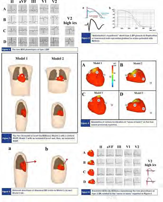
CONGRESSO
       NAZIONALE

     AIAC                                                                                                                          Bologna
                                                                                                                                   10-12 Marzo 2016

02
ARRHYTHMOGENIC RIGHT VENTRICULAR CARDIOMYOPATHY (ARVC) AND MYOCARDITIS CAN COEXSIST?
A COMPARATIVE STUDY BEETWEEN CARDIAC MAGNETIC RESONANCE (CMR) AND ENDOMIOCARDIAL
BIOPSY
G. Vettor 1, M. Casella 1, A. Dello Russo 1, G.M. Fassini 1, S. Riva 1, M. Moltrasio 1, F. Tundo 1, B. Majocchi 1, E. Russo 1, E. Conte 1, D. Andreini 1, G.
Pontone 1, S. Conti 1, F. Pizzamiglio 1, M.A. Dessanai 1, V. Marino 1, V. Catto 1, C. Basso 2, G. Thiene 2, C. Tondo 1
1
  Cardiac Arrhythmia Center Centro Cardiologico Monzino, Milano, ITALY
2
  Cardiovascular Pathology Università di Padova, Padova, ITALY

Introduction: Recent studies noted and supposed the possible coexistence of ARVC and myocarditis, considering the frequent finding of fibro-fatty
infiltration and an inflammatory component at histology. Contrast-enhanced cardiac magnetic resonance (CMR) is the only imaging modality able to
depict fibro-fatty infiltration and fibrosis as late gadolinium enhancement (LGE) deposition. The endomiocardial biopsy (EBM) plays an important role in
the definitive diagnosis of ARVC. The aim of our study was to investigate by CMR and EBM the possible coexistence of two pathologies.

Methods: From 2010 to October 2015, 62 patients were enrolled and underwent a CMR. For each patient a right ventricular (RV) mapping was
performed identifying the scar areas in the bipolar-unipolar maps. We obtained the right ventricular samples trough the right femoral vein via a
disposable biotome . All the samples obtained were guided by the electroanatomical voltage mapping.

Results: EBM was performed in 62 patients. In 7, 4 % (= 12) cases the biopsy was non diagnostic due to a sampling error. In 3% (=6) of patients
resulted aspecific. In the remaining 44 patients the biopsy showed the presence of an adipose infiltration with fibrosis with a diagnosis of ARVC in 18
(7,9%) patients, in 10 (4,4%) an histological inflammatory infiltrate as a myocarditis. In 16 patients (7%) the biopsy shows both a fatty infiltration and
an inflammatory component. In all cases of ARVC in the intraventricular septum and RV anterolateral wall LGE was noted. In none of patients, with
a no istopatological diagnosis of ARVC was noted fibrofatty-infiltration at CMR. In all patients with a istopatological myocarditis the LGE pattern was
midwall patchy. In remaining patients with a coexistence of ARVC and an inflammatory component the CMR presented only diagnostic criteria for
ARVC/D, and no suspicion of myocarditis LGE pattern.

Conclusion: In our experience in the most cases there is a concordance between the CMR and EBM in terms of scar areas-LGE and fibrosis in the
istopathological finding. Many cases of ARVC could be present also a inflammatory component mimicking a myocarditis, but the CMR was not to able
with accuracy to discriminate the inflammatory component in cases of coexistence of two pathologies.

                                                                            44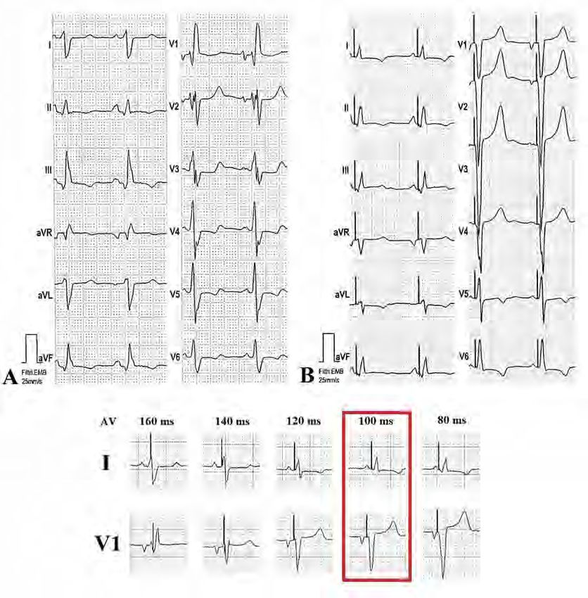
CONGRESSO
       NAZIONALE

     AIAC                                                                                                                         Bologna
                                                                                                                                  10-12 Marzo 2016

03
IMPIANTO DI ICD SOTTOCUTANEO IN PAZIENTE CON TRASPOSIZIONE DELLE GRANDI ARTERIE, DIFETTO
INTERVENTRICOLARE MUSCOLARE, DESTROCARDIA
R. Valecce, M. Palella, V. Bonfantino, C. Petrillo, M. Cuonzo, C. D’Agostino
Ospedale Di Venere, Bari, ITALY

Introduzione: La morte improvvisa (MI) è la principale causa di decesso in soggetti affetti da cardiopatie congenite complesse. La società europea
di cardiologia (ESC) ha elaborato indicazioni per l’impiego del defibrillatore sottocutaneo (S-ICD), all’interno delle Linee Guida 2015 per la prevenzione
della MI, attribuendogli indicazioni di classe IIA e di classe IIB. L’S-ICD appare proponibile soprattutto nei soggetti con anomalie del sistema venoso
e/o con cardiopatie congenite complesse che possono impedire o rendere rischioso un approccio transvenoso. L’S-ICD viene tipicamente impiantato
in regione toracica laterale sinistra, connesso ad un singolo elettrodo per sensing e defibrillazione, posizionato nel sottocute parallelamente alla linea
medio sternale. Esistono pochi lavori in letteratura sull’impianto dell’S-ICD in posizioni non standard.

Caso Clinico: Donna di 54 anni affetta da cardiopatia complessa: destrocardia in situs solitus, trasposizione delle grandi arterie, difetto interatriale ed
interventricolare ampio, stenosi valvolare polmonare severa. All’età di 2 anni veniva sottoposta ad intervento di shunt succlavio – polmonare. All’età di
16 anni veniva sottoposta, in sternotomia, a posizionamento di tubo di Dacron di 6 mm tra l’aorta ascendente ed il tronco polmonare successivamente
allungato di 1 cm per trombosi dello shunt. All’età di 34 anni le veniva posta diagnosi di sclerosi multipla a decorso recidivante-remittente. All’età di
45 anni ricovero per meningite batterica ed episodio di fibrillazione atriale cardiovertito elettricamente. Giungeva alla nostra osservazione in shock
e tachicardia ventricolare (TV) sostenuta, regredita mediante DC shock transtoracico a 200J. Veniva sottoposta ad ecocardiogramma, RX torace, e
angio TC che evidenziava occlusione dello shunt aorto polmonare. La SPO2 in O2 terapia a 4 l/min era 86-90%, con riduzione in decubito supino.
Dopo una accurata valutazione del nostro team circa i rischi correlati al posizionamento di un ICD con l’introduzione di un elettrocatere trasvenoso,
nonché le anomalie anatomiche cardiache e vascolari che avrebbero potuto rendere meno efficace la defibrillazione, si poneva indicazione ad impianto
di S-ICD. L’elettrocardiogramma mostrava un ritmo sinusale ed un blocco di branca destra. Lo strumento di screening ECG Boston Scientific è stato
utilizzato per determinare l’eleggibilità per impianto di S-ICD in posizioni differenti. Veniva impiantato pertanto un Boston Scientific Emblem S-ICD, con
posizionamento del generatore in regione toracica laterale destra tra il V ed il VI spazio intercostale ed il coil immediatamente alla destra dello sterno.
In considerazione delle condizioni clinico-emodinamiche della paziente, si è soprassieduto alla esecuzione del test di defibrillaizone.

Conclusioni: L’indicazione ad impianto di ICD nei pazienti affetti da cardiopatia congenita è aumentato negli ultimi decenni. Tuttavia, il posizionamento
di sistemi transvenosi ed epicardici in questa popolazione di pazienti non è scevra da rischi a medio-lungo termine. L’incidenza di fratture di
elettrocatetere, shock inappropriati, occlusione venosa ed infezioni sembra essere maggiore fino al 30% in pazienti affetti da cardiopatie congenite.
Abbiamo descritto l’impianto, sul lato destro del torace, di S-ICD in una giovane adulta affetta da cardiopatia congenita e destrocardia, registrando
adeguati valori di impedenza e sensing ed ottima morfologia dei QRS nei 3 vettori. Pertanto, il posizionamento di un S-ICD può fornire una ottima
alternativa ai sistemi epicardici o transvenosi in un gruppo selezionato di questi pazienti.

                                                                               45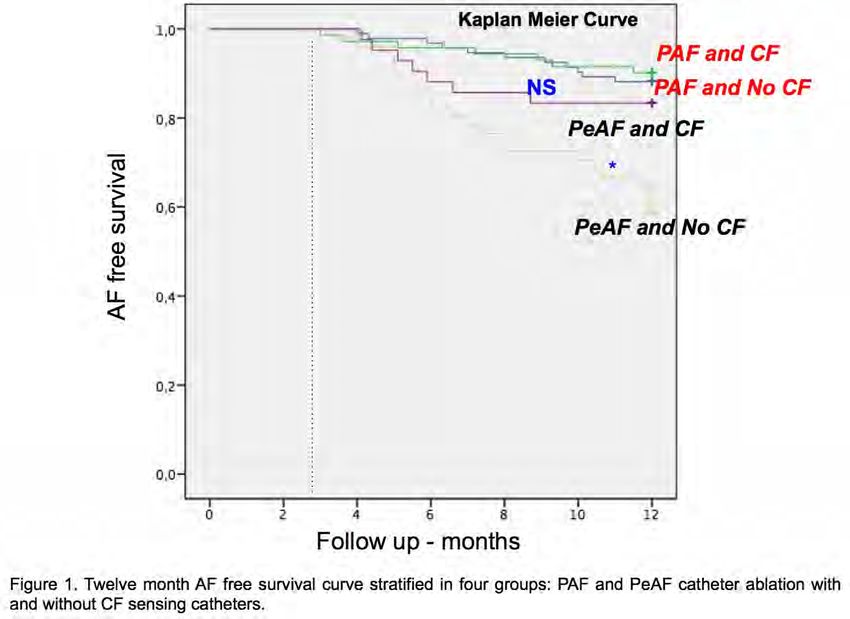
CONGRESSO
       NAZIONALE

     AIAC                                                                                                                              Bologna
                                                                                                                                       10-12 Marzo 2016

04
SICUREZZA DELLA CARDIOVERSIONE ELETTRICA DELLA FIBRILLAZIONE ATRIALE NEI PAZIENTI IN
TRATTAMENTO CON NUOVI ANTICOAGULANTI ORALI
M. Resta, L. De Gennaro, C. Campanella, L. Mancini, C. Borrelli, D. De Laura, G. Citarelli, D. Cavallari, F. Buquicchio, F. Corriero, P. Caldarola
Cardiologia Utic Ospedale San Paolo, Bari, ITALY

Introduzione: L’uso dei nuovi anticoagulanti orali (NAO) nella profilassi del tromboembolismo nei soggetti sottoposti a cardioversione elettrica (CVE) è
di relativa recente introduzione nella pratica clinica quotidiana. Abbiamo pertanto valutato la sicurezza di tale profilassi in un registro real-life di soggetti
sottoposti a CVE di fibrillazione atriale (FA), sia con protocollo accelerato che in modalità programmata, sia con che senza esecuzione preliminare di
ecocardiogramma trans esofageo (TEE).

Metodo: sono stati valutati 124 pazienti consecutivi sottoposti a CVE di FA: 40 sono stati sottoposti a CVE programmata dopo tre settimane di
terapia anticoagulante orale con NAO (50% con dabigatran, 25% con rivaroxaban e 25% con apixaban), 60 a CVE programmata dopo 3 settimane dal
raggiungimento di valori di INR compresi tra 2 e 3 con antagonisti di vitamina K (VKA). I pazienti sono stati sottoposti a TEE nel caso di INR non a target
o di dubbi sulla compliance alla terapia con i NAO. 24 pazienti, ricoverati per FA associata a sintomi di scompenso cardiaco o di ischemia, sono stati
sottoposti a CVE accelerata (in quest’ultimo gruppo 16 pazienti sono stati sottoposti a CVE dopo 24-48 ore dalla somministrazione dei NAO e TEE, 8
dopo somministrazione di eparine a basso peso molecolare e TEE). Sono stati quindi valutati gli eventi a breve termine (7 giorni) quali ictus, emorragie.

Risultati: nel gruppo di pazienti sottoposti a CVE programmata 72% dei pazienti era di genere maschile, età media 66 anni, nel 76% si trattava di
primo episodio, nel 20% avevano già eseguito una CVE,CHA2DS2VAScore medio era 2±1,3, l’HAS BLED medio era 1,17±0,64. Tra le comorbilità
la più rappresentata è stata l’ipertensione arteriosa (68%), seguita da diabete (16%), cardiopatia ischemica(16%)e distiroidismo (8%); nei pazienti
sottoposti a CVE accelerata 74% eranoipertesi, 30% diabetici, 26% affetti da cardiopatia ischemica, 8% con distiroidismo; nel 55% si trattava di primo
episodio, nel 9% si trattava di flutter atriale, il CHA2DS2VAScore era di 3,17±1,44, HAS BLED 1,48±0,85. Nel primo gruppo l’ecocardiogramma trans
esofageo (TEE) è stato eseguito nel 43% dei pazienti, in particolare nel 60% dei pazienti in terapia con NAO, nel 15% di pazienti con VKA con INR a
target e nel 100% dei pazienti in terapia con VKA con INR non a target. La velocità media in auricola era di 40 cm/sec nei soggetti sottoposti a CVE
elettiva, in nessun caso sono stati riscontrati trombi o sludge che controindicavano la CVE, in 4 casi è stato riscontrato eco-contrasto spontaneo (uno
di questi assumeva apixaban 5 mg, UN ALTRO Rivaroxaban 20 mg, uno VKA con INR a target, uno con VKA con INR non a target). Nel secondo gruppo
la velocità media in auricola era di 35±7.07, in 2 pazienti non è stata eseguita CVE per riscontro dimarcato effetto sludge

                                                                               46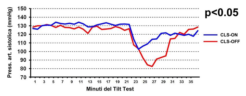
CONGRESSO
       NAZIONALE

     AIAC                                                                                                                           Bologna
                                                                                                                                    10-12 Marzo 2016

05
ABLAZIONE TRANSCATETERE NELLA POPOLAZIONE PEDIATRICA: MINIMIZZARE L’ESPOSIZIONE
RADIOSCOPICA È POSSIBILE
P. Ferrari, F. Giofre’, P. De Filippo
Azienda Ospedaliera Papa Giovanni XXIII, Bergamo, ITALY

Razionale: L’ablazione transcatetere (aTC) delle aritmie si basa sull’utilizzo di fluoroscopia. Il bambino è maggiormente radiosensibile per la crescita
cellulare e per la maggior aspettativa di vita. I sistemi di mappaggio elettroanatomici (MAP) e l’ottimizzazione del settaggio degli apparati di fluoroscopia
si sono dimostrati efficaci nel ridurre la dose di radiazioni ionizzanti.

Metodi: questo è uno studio monocentrico, retrospettivo. Tutti i pazienti di età inferiore ai 18 anni sottoposti ad ablazione transcatetere nel nostro
Centro da Giugno 2013 a Novembre 2015 sono stati rivisti e divisi in due gruppi. Questo studio confronta un gruppo A di pazienti con fluoroscopia
standard (SD) + MAP con un gruppo B dove è stato adottato un protocollo di riduzione della frame rates e di bassa fluoro dose basato sul peso del
paziente (LD) +MAP. Obiettivo dello studio era valutare la riduzione della dose radioscopica al paziente (DAP).

Risultati: 56 pazienti consecutivi sottoposti ad aTC sono stati inclusi. Nel gruppo A l’età media era 13,5 anni, il peso 53 Kg ed il BMI 20 m2 . Nel
gruppo B l’età media era 10,9 anni, il peso 41,9 Kg ed il BMI 18,4 m2 . Non vi era differenza statisticamente significativa nel BMI fra i due gruppi. La
distribuzione delle tachicardie era bilanciata nei due gruppi. Nel gruppo A: 53% AVRT, 42% AVNRT e 5% altre. Nel gruppo B: 54% AVRT, 40% AVNRT
e 6% altre.
Nel gruppo B la DAP è stata ridotta di un terzo rispetto al gruppo A (151,9 vs 533,5 µGym2, p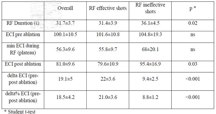
CONGRESSO
       NAZIONALE

     AIAC                                                                                                                          Bologna
                                                                                                                                   10-12 Marzo 2016

06
IL SEGNO DELLA “RADICE QUADRATA” NELLA REGISTRAZIONE BIPOLARE INDICA L’ORIGINE IATROGENA
DELLE EXTRASISTOLI DURANTE STUDIO ELETTROFISIOLOGICO
G. Epicoco 1, H. Ali 1, A. Sorgente 2, S. Foresti 1, G. De Ambroggi 1, A. Fundaliotis 1, P. Lupo 1, R. Cappato 1
1
  Humanitas Gavazzeni, Bergamo, ITALY
2
  Cleveland Clinic Abu Dhabi, Abudhabi, UNITED ARAB EMIRATES

Introduzione: In corso di studio elettrofisiologico vengono frequentemente indotte extrasistoli iatrogene causate dalla stimolazione meccanica della
punta dell’elettrocatetere sulla superficie endocardica. La comparsa delle extrasistoli consensualmente al movimento dell’elettrocatetere e il differente
aspetto elettrocardiografico rispetto ai battiti spontanei, solitamente, caratterizzano l’origine meccanica. Tuttavia, durante il mappaggio di aritmie
focali, differenziare i battiti spontanei da quelli iatrogeni non è sempre agevole. Considerando l’elettrogramma (EGM) bipolare come “mini-unipolare”
quando l’impulso elettrico origina da una porzione di miocardio localizzata al di sotto della punta del catetere mappante, la morfologia del segnale
bipolare può aiutare a riconoscere le extrasistoli iatrogene. Scopo dello studio è stato dimostrare che la presenza di un pattern a “radice quadrata”
dell’EGM bipolare identifica le extrasistoli iatrogene.

Metodi: Durante studio elettrofisiologico sono stati registrati i segnali endocavitari di 63 extrasistoli atriali e di 64 extrasistoli ventricolari indotte
meccanicamente mediante movimento dell’elettrocatetere ablatore in 10 pazienti con cuore strutturalmente normale. Sono stati analizzati gli EGM
unipolari (0.5-500 Hz) e bipolari (30-500Hz) registrati in contemporanea in corrispondenza dei battiti iatrogeni. Il pattern a “radice quadrata” è stato
definito come la presenza di una rapida deflessione negativa nei primi 20 ms dell’EGM bipolare seguita da una componente positiva o isodifasica.

Risultati: Un EGM unipolare con morfologia QS è stato registrato in corrispondenza di tutte le extrasistoli indotte. Il picco dell’EGM bipolare ha
anticipato l’inizio dell’onda P o del QRS extrasistolico sull’ECG di superficie rispettivamente di 44 ± 14 ms o 34 ± 7 ms. Il voltaggio del segnale
bipolare delle extrasistoli atriali e ventricolari è stato, rispettivamente, 1,4 ± 0,9 mV e 5,8 ± 1,6 mV. Significativamente, un pattern a “radice quadrata”
è stato rilevato in 63/64 extrasistoli atriali (98%) e in 62/63 extrasistoli ventricolari (98%).

Conclusioni: Un pattern a “radice quadrata” dell’EGM bipolare può essere considerato un indice affidabile nell’identificare l’origine meccanica delle
extrasistoli atriali e ventricolari.

                                                                              48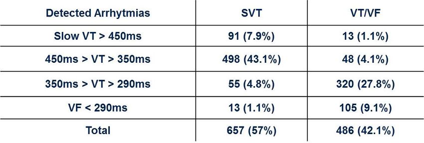
CONGRESSO
       NAZIONALE

     AIAC                                                                                                                            Bologna
                                                                                                                                     10-12 Marzo 2016

07
SAFETY AND EFFICACY OF RANOLAZINE FOR THE PREVENTION OF DRUG-REFRACTORY VENTRICULAR
ARRHYTHMIAS AND RECURRENT ICD INTERVENTIONS
F. Salghetti, A.Curnis, N. Dasseni, E. Sciatti, M. Cerini, A. Lipari, F. Vassanelli, L. Inama, A. D’Aloia, E. Vizzardi, L.Bontempi
Division of Cardiology, University of Brescia, Spedali Civili, Brescia,ITALY

Background: There are limited options for patients who present with antiarrhythmic-drug (AAD)-refractory ventricular arrhythmias (VA) and recurrent
implantable cardioverter defibrillator (ICD) interventions. Ranolazine inhibits the late Na current and is an antiischemic and antianginal agent approved
in 2006 for use in stable angina pectoris. Several experimental and preclinical data (in vivo ad in vitro) support additional antiarrhythmic properties of
ranolazine, in both atrial and ventricular myocardium. However the clinical effect and the safety of ranolazine when added to standard anti-arrhythmic
drugs (AADs) treatment for the suppression of drug-refractory ventricular arrhythmias, is currently uncertain and no randomized control clinical trials
have been published yet.

Objective: To evaluate the safety and efficacy of ranolazine as antiarrhythmic agent in implantable cardioverter defibrillator (ICD) recipients with
recurrent sustained ventricular arrhythmias refractory to optimized conventional AADs/heart failure treatment and to assess any differences in
effectiveness as antiarrhythmic agent in ischemic patients (CAD+) compared to non-ischemic patients (CAD-).

Patients and Methods: In this single-center case-series, 17 patients with ischemic or non-ischemic cardiomyopathy, with existing devices (ICD/
CRT-D), who have experienced VT/VF appropriately treated with ICD therapy (Anti-Tachycardic Pacing or DC shock) or had several NSVTs lasting at
least 10 beats, were enrolled (Arrhythmia Group). Because of refractory VT or intolerance to the medications employed, alternative methods to control
the VA were sought. Each patient was on a class III antiarrhythmic and/or beta-blockers optimized medication and Ranolazine was added. 12 ICD
recipients who, in the same period of time, began Ranolazine because of recurrent stable angina, but without any major VA at the time of enrollment,
were also considered (Angina Group) . The study endpoints was the number of Non sustained ventricular tachycardia (NSVT), Ventricular Tachycardia
(VT), Ventricular Fibrillation (VF), Anti-tachycardic pacing (ATP) or DC shock delivery at 1 week, 6, 12 and 18 months from the starting dose.

Results: 17 patients in the Arrhythmia Group and 12 patients in the Angina Group were enrolled. The average age was 67 ± 12 years and 72 ± 8
years respectively. In the Arrhythmia group, 6 (35.3%) patients had ischemic cardiomiopathy (CAD+) with average Ejection Fraction (EF) 32.4% ±
5.1 ; of those with non-ischemic cardiomiopathy (CAD-), 10 (58.8%) had idiopatic cardiomiopathy with average EF 30.8% ± 4.6, and 1 (5.9%) had
arrhythmogenic right ventricular cardiomyopathy/dysplasia (ARVC) with preserved EF. Over 12 ± 6 months, in the Arrhythmias Group Ranolazine the
presence of NSVT (p 0.021), VT (p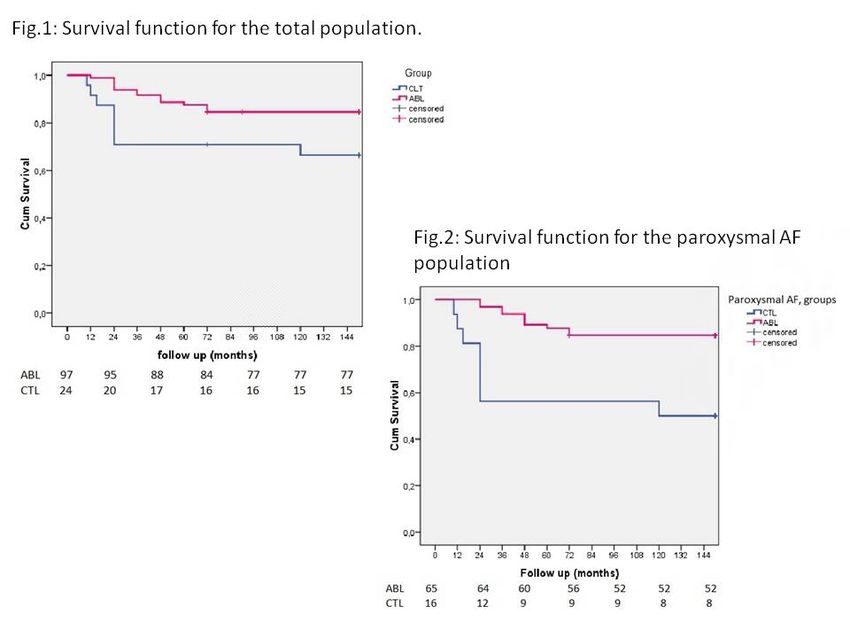
CONGRESSO
NAZIONALE
AIAC Bologna
10-12 Marzo 2016
08
A THREE-DIMENSIONAL ELECTROPHYSIOLOGICAL COMPUTED MODEL OF ST SEGMENT ABNORMALITIES
IN TYPE 1 BRUGADA PATTERN: THE KEY ROLE OF RIGHT VENTRICULAR OUTFLOW TRACT ORIENTATION
IN THE THORAX
P. Crea, G. Picciolo, F. Luzza, G. Oreto
Dipartimento di Medicina Clinica e Sperimentale, Università di Messina, Messina, ITALY
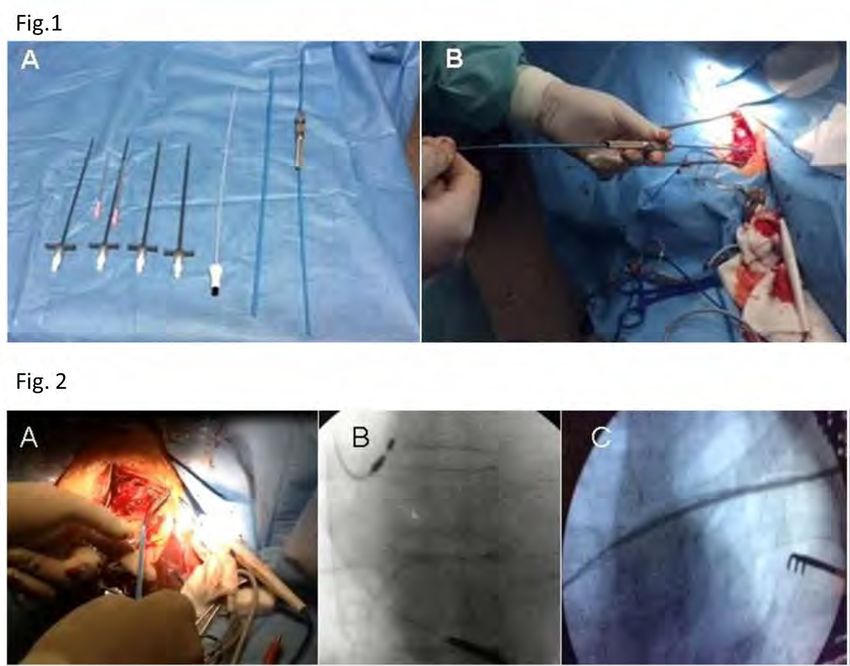
A recent study, conducted by our group, reported that slightly less than one half of patients with type 1 Brugada Pattern (BP) show a definite ST
segment depression (>0.1 mV with duration > 0.08 s) in the inferior leads. Thus, 4 distinct ST abnormalities phenotypes were recognized in Type 1
BP: Phenotype A) Type 1 BP diagnosis in V1-V2 at 4th ics, no ST segment depression in inferior leads (Fig. 1A); Phenotype B) Type 1 BP diagnosis in
V1-V2 at 4th ics, ST segment depression in inferior leads ( > 0.1 mV with duration >0.08 s) (Fig. 1B); Phenotype C) Type 1 BP diagnosis at high ics
(3rd or 2nd), no ST segment depression in inferior leads (Fig. 1C); Phenotype D) Type 1 BP diagnosis at high ics (3rd or 2nd), ST segment depression
in inferior leads ( > 0.1 mV with duration > 0.08 s) (Fig. 1D). Several studies found a correlation between the right ventricular outflow tract (RVOT)
anatomical location and the leads in which a diagnostic type 1 Brugada pattern could be observed. We hypothesized the key role of orientation of right
ventricular outflow tract in the chest, particularly the inclination of anterior wall compared to the sternum, contributing to the determination of these
various ECG phenotypes. In order to assess our hypothesis we used an interactive simulation program able to assess the relationship between the
electric current sources of the heart and the resulting electrocardiographic signals on the body surface (ECGSIM vers. 3.0.0 ). This computed model
supports the strict relationship between ST segment depression in the inferior leads and the ST segment elevation in right precordial leads, typical of
type 1 BP. A horizontal RVOT, in fact, gives raise to abnormal BP vector directed both superiorly and anteriorly, explaining, at the same time, typical BP
appearance in right precordial leads and ST segment depression in the inferior leads. Analysis of the inferior leads could be useful especially in patients
with no clear type 1 BP with V1-V2 at the 4th ics suggesting the need to record upper right precordial leads. Cardiac imaging, such as computed
tomography and magnetic resonance, or perhaps echocardiography, could find a further validation of this hypothesis. Further studies needs to assess
a possible prognostic role of various ECG phenotypes of type 1 BP.
50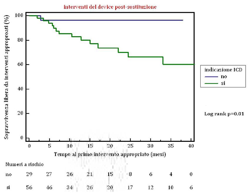
CONGRESSO
       NAZIONALE

     AIAC                                                                                                                      Bologna
                                                                                                                               10-12 Marzo 2016
GIOVEDI’ 10 MARZO 2016

13.30 - 14.00
AREA POSTER
SESSIONE E-POSTER 2- ABLAZIONE DELLE ARITMIE E CHIUSURA DELL’ AURICOLA

01
CONTACT FORCE VS NON-CONTACT FORCE GUIDED SURROUND FLOW CATHETER ABLATION OF ATRIAL
FIBRILLATION IN A HIGH VOLUME CENTER.
D. Dugo 1,2, S. Bordignon 1, L. Perrotta 1, F. Bologna 1, F.K. Weise 1, A. Fürnkranz 1, B. Schmidt 1, J.K.R. Chun 1
1
  Ccb - Markuskrankenhaus, Francoforte sul Meno, GERMANY
2
  P.O. Ferrarotto - U.O. Cardiologia, Catania, ITALY

Purpose: Radiofrequency current (RFC) catheter ablation is an effective treatment in symptomatic atrial fibrillation (AF). To improve RFC lesions novel
catheters have been introduced combining improved catheter tip cooling (ThermoCool surround flow, TC-SF, Biosense Webster) and local contact force
information. We therefore aimed to study procedural safety and efficacy of these new catheters.

Methods: Our data base comprises 3000 AF ablation procedures (May 2010-November 2015). All AF ablation procedures performed between July
2014 and May 2015 using the novel contact force + surround flow catheter (group A: SmartTouch SF, ST-SF, Biosense Webster) and CARTO 3D
mapping system were retrospectively analyzed. Only patients with a minimum follow up of 6 months were included in the study. A matched control
group consisted of surround flow cooled tip ablation without contact force (group B: TC-SF, Biosense Webster). Ablation energy was limited to 30W, 8
mL/min (posterior LA) and 40W, 15 mL/min (anterior LA) respectively. Procedural success, complications and outcome were analyzed.

Results: A total of 52 patients was identified. Group A (ST-SF) and B (TC-SF) consisted of 26 pts, respectively 54% and 50% males, mean age 66
± 9 and 67 ± 10 years, LA 40 ± 7 and 42 ± 4 mm, 96% and 81% PAF; for all p=n.s.) Mean procedural and fluoroscopy times were significantly
shorter in group B (group A: 98 ± 32 min and 11 ± 7 min vs group B: 78 ± 31 and 7 ± 3 min; for both p< 0.05). Complete PV isolation proven with
a spiral catheter was obtained in all patients (100%). No major complications occurred in both groups (tamponade: n=0, stroke: n=0, AE fistula: n=0);
regarding minor complications, no differences were reported (vascular access: n=1 in group A vs n=0 in group B; p=ns.). Stable sinus rhythm was
observed without differences in both groups (group A: 77%, mean FU: 305 ± 104 days vs. group B 73%, mean FU: 255 ± 84 days, for both p=n.s.).

Conclusion: Catheter ablation using contact force information appears to be safe in AF ablation. However, the addition of contact force information
in a high volume center did not improve procedural and clinical outcome.

                                                                             51
CONGRESSO
      NAZIONALE

     AIAC                                                                                                                   Bologna
                                                                                                                            10-12 Marzo 2016

02
CATHETER ABLATION OF ATRIAL FIBRILLATION USING THE SECOND-GENERATION CRYOBALLOON VERSUS
OPEN-IRRIGATED CONTACT-SENSING RADIOFREQUENCY: 12 MONTHS OUTCOME BASED ON INSERTABLE
CARDIAC MONITOR
G. Sirico, S. Panigada, D. Sacchetta, V. De Sanctis, M. Mantica
Istituto Clinico Sant’Ambogio, Milano, ITALY

Introduction: There are limited comparative data on catheter ablation (CA) of atrial fibrillation (AF) using the second-generation cryoballoon (CB-
2) versus point-by-point contact-force guided radiofrequency (RFCF). Patients’ symptoms, electrocardiograms or 24-hour Holter recordings often
underestimate AF relapses after CA when compared with insertable cardiac monitoring (ICM). We here report results of AF CA using CB-2 versus RFCF
based on ICM and continuous home monitoring.

Methods: From January 2014 to January 2015, 76 patients affected by paroxysmal AF underwent first pulmonary vein isolation (PVI) using CB-2 (n
= 25) or RFCF (n = 51) in a non-randomized fashion. A Reveal Linq ICMTM was implanted following CA in 9 and 8 patients who underwent CB-2 and
RFCF CA, respectively. Those ICM patients were connected with MyCareLinqTM home monitoring system offering daily automatically wireless data
transmissions. ICM recorded the amount of AF per last follow-up year (AF burden). AF recurrence was considered as any episode of AF, atrial flutter
or atrial tachycardia lasting at least 30 seconds after blanking period with or without symptoms.

Results: PVI was successfully obtained in all patients. On ICM, AF burden was 0% in 8 out of 9 (88,9%) CB-2 and in 7 out of 8 RFCF (87,5%) patients,
at 1 year follow up (p=0.567). In CB-2 group one patient showed AF burden of 11.9% (after redo procedure using RF), while in RFCF group one patient
showed AF burden of 0,6% (after single CA). All patients were free from antiarrhythmic drugs at the end of blanking period. AF related symptoms had
the same prevalence in both groups, CB-2 and RFCF (p=0.235).

Conclusions: ICM offers greater efficacy in AF recurrence monitoring after CA compared with non invasive recorders. Based on ICM, CB-2 and RFCF
supported CA of paroxysmal AF showed similar 12 months outcome after single procedure.

                                                                        52
CONGRESSO
NAZIONALE
AIAC Bologna
10-12 Marzo 2016
03
CATHETER ABLATION IMPROVES SURVIVAL IN PAROXYSMAL ATRIAL FIBRILLATION PATIENTS. 12-YEAR
FOLLOW-UP OF A PROSPECTIVE, MULTI-CENTER, RANDOMIZED, CONTROLLED STUDY (THE CACAF
STUDY).
S. Ferretto 1, G. Senatore 2, L. De Michieli 1, A. De Simone 3, C. Amellone 2, V. La Rocca 3, M. Giuggia 2, D. Corrado 1, F. Zoppo 4, G. Stabile 5, E. Bertaglia 1
1
Dipartimento di Scienze Cardiologiche, Toraciche e Vascolari, Università di Padova, Padova, ITALY
2
Ospedale Civile, Ciriè, Torino, ITALY
3
Casa di Cura San Michele, Maddaloni, Caserta, ITALY
4
Ospedale Civile, Mirano, Venezia, ITALY
5
Clinica Mediterranea, Napoli, Napoli, ITALY
Introduction: Until now, randomized and controlled study have reported the effect of catheter ablation (CA) for atrial fibrillation (AF) over a follow-up
of 12-24 months. None study has reported the effect of CA on progression to permanent AF beyond 10 years. This is a multicenter, randomized,
controlled study which reports the results of the extended follow-up (12 years) of the Catheter Ablation for the Cure of Atrial Fibrillation (CACAF) Study.
Aim: To determine the effect of CA on sinus rhythm maintenance and cumulative survival in patients with AF.
Methods: We performed an “on-treatment” analysis of the 137 patients enrolled between 01st February 2001 and 30th June 2003 in the CACAF
Study. Study population was composed by patients affected by paroxysmal or persistent AF, divided in two groups on the basis of the received
treatment: CA + antiarrhythmic drugs (ABL group) vs antiarrhythmic drugs alone (CTL group). Patients underwent an in-office visit or phone interview,
and were invited to repeat a 12-lead ECG 144±3 months after enrollment.
Results: One hundred twenty one patients were available for analysis (16/137, 11.7% of CACAF patients were lost from follow up): 97 in the ABL
group and 24 in the CTL group. At the 144-month follow-up examination, 54/97 (55.7%, 95% CI 45.8-65.2%) ABL patients and 3/24 (12.5%, 95% CI
4.3-31.0%) CTL patients were alive and in sinus rhythm (pCONGRESSO
NAZIONALE
AIAC Bologna
10-12 Marzo 2016
04
FOLLOW-UP A 24 MESI DOPO CRIOABLAZIONE DELLE VENE POLMONARI NEI PAZIENTI CON FIBRILLAZIONE
ATRIALE PAROSSISTICA. CONFRONTO TRA LE DUE GENERAZIONI DI CRIOPALLONI.
A. Colella
Aritmologia, Careggi, Firenze, ITALY
Introduzione: La crioablazione (Cryoballon, Medtronic, USA) e’ risultata efficace per isolare le vene polmonari con una singola applicazione di energia.
Una seconda generazione di criopalloni (Advance, Medtronic) e’ stata costruita per avere una piu’ ampia zona ghiacciata di contatto con il tessuto
atriale e per ottenere una lesione circonferenziale piu’ uniforme e delimitata. In questo studio vengono messi a confronto i due tipi di criopalloni (CB-1
e CB-2) e valutato sia la sicurezza che l’outcome a lungo termine dell’efficacia dell’isolamento delle vene polmonari.
Materiale e Metodi: Sono stati analizzati i dati dei 79/106 pazienti consecutivi affetti da FA parossistica, con assenza di alterazioni strutturali atriali
e patologie cardiovascolari associate, che sono arrivati al termine di un follow-up di 24 mesi. I pazienti sottoposti a CRIOABLAZIONE delle vene
polmonari sono stati suddivisi in due gruppi (CB-1 N=49; CB-2 N=30 ). La disconnessione elettrica delle vene polmonari, nella nostra procedura di
ablazione, viene da sempre validata dal catetere circolare Achieve (Medtronic, USA) inserito nel lume del criopallone.
Risultati: Dopo un “blanking period” di 3 mesi, una singola procedura di crioablazione e senza terapia farmacologia associata al termine del follow-up,
sono senza FA il 78% (38/49 pz) del gruppo CB-1 e l’80% (24/30 pz) del gruppo CB-2 (p=0.39). Con la seconda generazione di criopalloni si ottiene
una significativa riduzione, delle applicazioni sulle vene polmonari sinistre (CB-1=1,84±0,37 vs CB-2=1,47±0,50; pCONGRESSO
NAZIONALE
AIAC Bologna
10-12 Marzo 2016
05
USE OF ELECTRICAL COUPLING INDEX IN TYPICAL ATRIAL FLUTTER ABLATION
M. Maines, D. Catanzariti, C. Angheben, M. Del Greco
Ospedale Santa Maria del Carmine, Rovereto, ITALY
Introduction: A new generation ablation system with an irrigated ablation catheter in conjunction with an advanced electro-anatomic mapping and
navigation system allows the evaluation of the Electrical Coupling Index (ECI), an indication of tip-to-tissue contact. Aim of our study was to evaluate
if this index could also give an indication about ablation lesion efficacy.
Methods: In patients undergoing typical right atrial flutter ablation, we compared the values of the ECI before, during (at the plateau) and after isthmus
ablation. Permanent tissue damage or ablation lesion efficacy was defined as the reduction in the local potential > 90% or as potential split in 2
separate signals. In absence of these endpoints, lesions were deemed ineffective.
Results: 15 consecutive patients (11 males, age 69.3±11.4 years) with history of typical atrial flutter underwent an ablation with Contact™ Therapy™
Cool Path™ Cardiac Ablation System in conjunction with EnSite™ Velocity Contact™ technology between Sep 2012 and Aug 2013. Target site for
ablation was the isthmus between the inferior vena cava and the tricuspid valve. All the procedures were successful, without complications. The
number of radiofrequency (RF) applications was 10.8±6.7 (range 6-28) and RF time was 330.3±177.5s. ECI values are reported in the table:
RF effective applications needed less time and the ECI post-ablation was inferior compared to ineffective RF applications. The absolute and percentage
ECI variations (pre-post ablation) were significantly greater when applications were effective (pCONGRESSO
NAZIONALE
AIAC Bologna
10-12 Marzo 2016
06
IS A SYSTEMATIC USE OF MAPPING SYSTEMS DURING CATHETER ABLATION PROCEDURES IN CHILDREN
AND TEENAGERS COST EFFECTIVE? A SNAPSHOT OF OUR EXPERIENCE
F. Guarracini 1, M. Marini 1, M. Del Greco 2, D. Ravanelli 3, A. Cima 2, A. Coser 1, G. Porcedda 4, A. Valentini 3, R. Bonmassari 1
1
Department of Cardiology, S. Chiara Hospital, Trento, ITALY
2
Department of Cardiology, S. Maria del Carmine Hospital, Rovereto (TN), ITALY
3
Department of Physics, S. Chiara Hospital, Trento, ITALY
4
Department of Pediatrics, Mayer Hospital, Firenze, ITALY
Introduction: Standard imaging during catheter ablation (CA) uses fluoroscopy. The aim of this study was to evaluate the cost effectiveness of an
extended use of mapping systems (MS) during paediatric CA in an adult EP Lab.
Methods: This study is a retrospective analysis that includes consecutive young patients (58 pts, aged between 8-18) who underwent CA from
March 2005 to February 2015. We compare the fluoroscopy data of two groups of pts: group I, pts who underwent CA from 2005 to 2008 using only
fluoroscopy and group II, pts who underwent CA from 2008 to 2015 performed also using MSs.
Results: The use of a MS during CA resulted in a reduction of the fluoroscopy time for pts in Group II by comparison with pts in Group I and the
difference between the two groups in median effective dose was 2.8 mSv (3.04 mSv (95% CI [1.22-6.89]) in Group I and 0.25 mSv (95% CI
[0.00-0.60]) (MW-test, P < 0.05) in Group II). If we consider the man-sievert monetary value, i.e. the monetary reference value of the avoided unit
of exposure (referred to by the ICRP), we can use this value to judge the cost-effectiveness of the use of MS during CA. However, the use of the
monetary value varies markedly between countries. There is a wide range from the lowest value adopted by UK (12.55 €/mSv) to the highest values
in Switzerland (2481.39 Times /mSv). For this study we referred to the value adopted in Netherland (453.78 Times /mSv). A further issue is that it is
recognized that the monetary value has to be adjusted for children and the accepted protection cost can be at least 3 times higher (37.65 Euro/mSv
UK, 1361.64 Euro/mSv in Netherland and 7444.17 Euro/mSv in Switzerland). Our extra cost of using a MS for CA is Times 2,500 per pt. It is evident
if we compare our cost with the man-sievert monetary value in the Netherland, it is cost-effective (1361.64 Euro/mSv*2.8 mSv= 3811.75 Euro/pt vs
Times 2,500 per pt).
Conclusions: The amount of X-ray exposure reduction reported in our “real-life” study makes a strong case for the daily use of a MS during CA and
it seems to be cost effective.
56CONGRESSO
NAZIONALE
AIAC Bologna
10-12 Marzo 2016
07
PERCUTANEOUS LAA OCCLUSION: ARE SPONTANEOUS ECHO CONTRAST (SEC) OR SLUDGE A
CONTRAINDICATION TO DEVICE IMPLANTATION?
G. Vettor, M.G. Fassini, S. Riva, A. Dello Russo, M. Casella, S. Conti, M. Pepi, C. Tondo, A.M. Maltagliati
Cardiac Arrhythmia Center Centro Cardiologico Monzino, Milano, ITALY
Introduction: Percutaneous closure of LAA is emerging as an alternative to pharmacological approach for thromboembolism prevention in patients
with non-valvular atrial fibrillation (AF). Exclusion of atrial thrombi is mandatory in patients’ candidates to percutaneous LAA occlusion. Multiplan
trans esophageal echocardiography (MTEE) is the gold standard for atrial thrombi visualization and allows identification of spontaneous echo contrast
(SEC) and sludge, defined as a dynamic, viscid layered echo density without a discrete mass. Patients with atrial fibrillation and SEC have a high risk
of thromboembolic events and death. We report our experience on procedural safety and clinical outcomes of patients with different degrees of SEC
candidate to percutaneous LAA closure
Methods: From March 2010 to July 2015 eighty patients with contraindication to anticoagulant therapy or recurrent cerebrovascular events despite
anticoagulant therapy were enrolled for LAA closure. MTEE was performed within 48 hours before the device implant. After the procedure, a clinical
and echocardiographic follow-up was arranged.
Results: Successful LAA closure was achieved in 72 cases (92,8%, 16 with sludge). In 3 patients (4,6%) periprocedural serious complication, not
related to the presence of severe SEC or sludge, were observed. During follow-up, MTEE showed thrombosis on the atrial surface of the device in 2
patients with sludge. No embolic or bleeding events were observed in patients with severe SEC or sludge.
Conclusion: the detection of very severe SEC with sludge features does not increase the incidence of periprocedural complications, especially of
thromboembolic events. Therefore SEC or sludge is not a contraindication to LAA closure. However during follow-up uncommonly thrombosis of the
device may occur and an accurate and closer monitoring is mandatory.
57CONGRESSO
NAZIONALE
AIAC Bologna
10-12 Marzo 2016
08
UTILITÀ DELL’ALGORITMO AUTOMATICO PASO NEL MAPPAGGIO CARTO E NELL’ABLAZIONE
DELL’EXTRASISTOLIA VENTRICOLARE
M. Casella, F. Pizzamiglio, S. Conti, M. Zucchetti, S. Arnoldi, E. Russo, G. Vettor, B. Majocchi, F. Tundo, M.A. Dessanai, V. Marino, M. Moltrasio, G. Fassini,
S. Riva, C. Carbucicchio, A. Dello Russo, C. Tondo
Cardiac Arrhythmia Research Centre, Centro Cardiologico Monzino Irccs, Milano, ITALY
Introduzione: L’ablazione con radiofrequenza è solitamente curativa nei confronti dell’extrasistolia ventricolare in cuore sano. Tuttavia, l’outcome
procedurale dipende dalla possibilità di registrare frequenti battiti extrasistolici durante lo studio. In caso di extrasistolia sporadica, il pace-mapping
rappresenta uno strumento utile per identificare il sito ottimale di ablazione, ma il confronto manuale dei tracciati è soggettivo e richiede spesso molto
tempo.
Metodi: Abbiamo arruolato 29 pazienti (età media 50±21 anni) senza malattia strutturale cardiaca che si presentavano con diversi tipi di extrasistolia
ventricolare. Tutti sono stati sottoposti a procedura ablativa utilizzando il software PaSo di analisi automatica del pace-mapping (Pace-Mapping
Software, Biosense Webster Inc.). La mappa di voltaggio in ritmo sinusale è stata effettuata in tutti i pazienti, mentre la mappa di attivazione è stata
effettuata solo in pazienti con extrasistolia frequente. Il pace-mapping è stato effettuato in sedi multiple e il PaSo è stato effettuato analizzando
ciascuna derivazione e calcolando sulle 12 derivazioni uno score di corrispondenza da 0 a 1.0. Sono stati considerati siti di ablazione adeguati solo i
siti con corrispondenza PaSo di almeno 0.8 in 12/12 derivazioni ECG.
Risultati: Durante la procedura i pazienti presentavano una frequenza variabile di battiti extrasistolici: 5 (17%) avevano un battito extrasistolico ogni
3 minuti, 16 (55%) un battito extrasistolico ogni minuto, 10 (34%) presentavano fasi di bigeminismo. La mappa di voltaggio in ritmo sinusale è stata
effettuata in tutti i pazienti (media dei punti acquisiti 194±110), mentre la mappa di attivazione è stata effettuata in 20 (69%) casi. Il pace-mapping
è stato effettuato in una media di 4.59 siti. Il tempo totale di mappaggio e pace-mapping è stato 56±32 minuti. L’ablazione è risultata efficace in 23
pazienti (79%), di cui 11 (48%) in ventricolo destro e 12 (52%) in ventricolo sinistro. I siti di ablazione efficace presentavano una corrispondenza PaSo
di 0.92±0.05 e una precocità media di 32±5 ms nei confronti dell’onset del QRS. La differenza tra i valori PaSo nei siti di ablazione efficace e i siti di
ablazione inefficace (0.64±0.05) è risultata statisticamente significativa (pCONGRESSO
NAZIONALE
AIAC Bologna
10-12 Marzo 2016
GIOVEDI’ 10 MARZO 2016
13.30 - 14.00
AREA POSTER
SESSIONE E-POSTER 3 - I DEVICE IN ARITMOLOGIA
01
DOWNGRADING FROM CRT-D TO CRT-P AT THE TIME OF BATTERY DEPLETION: PRELIMINARY RESULTS
FROM DECODE REGISTRY TRIAL
V. Zacà 1, M.L. Narducci 2, Q. Parisi 3, D. Potenza 4, F. Zanon 5, M. Manzo 6, G. Stabile 7, M. Iori 8, P. De Filippo 8, M. Zoni Berisso 8, A. Pierantozzi 8, A.
Bandini 8, L. Calò 8, P. Sabbatani 8, M. Zennaro 8, M.S. Argnani 8, E. Ammendola 8, F. Picariello 9, M. Malacrida 9, M. Biffi 10
1
Hospital S. Maria alle Scotte, Siena, ITALY
2
Catholic University of the Sacred Heart - Institute of Cardiology, Roma, ITALY
3
Catholic Univ of the Sacred Heart Cardiovascular Dept, Campobasso, ITALY
4
Casa Sollievo della Sofferenza, San Giovanni Rotondo (FG), ITALY
5
Ospedale S. Maria della Misericordia, Rovigo, ITALY
6
A.O. Ruggi D’Aragona, Salerno, ITALY
7
Clinica Mediterranea, Napoli, ITALY
8
Decode Registry Group, Milano, ITALY
9
Boston Scientific Italia, Milano, ITALY
10
Institute of Cardiology, University of Bologna, Policlinico S.Orsola-Malpighi, Bologna, ITALY
Introduction: Data from Madit-CRT showed that CRT-defibrillator (CRTD) patients (pts) who achieve ejection fraction (LVEF) normalization (>50%)
have very low absolute and relative risk of Ventricular Tachyarrhythmias (VTAs) and a favorable clinical course within 2.2 years of follow-up. These pts
could be considered for downgrade to CRT-pacemaker (CRTP) at the time of battery depletion if no VTAs have occurred.
Methods: The DECODE Registry enrolled consecutive pts who underwent CRTD replacement from 2013 to 2015 in 36 Italian centers. At the time
of replacement, clinical and echocardiographic findings were assessed and the number of appropriate therapies delivered by the replaced device or
pre-implantation history of VTAs was retrieved. The occurrence of appropriate ICD therapies was measured during follow-up.
Results: A total of 313 pts with complete data were considered for this analysis (age 70±10 years, male gender 73%, ischemic etiology 47%, NYHA
class I/II at replacement 64%, mean LVEF 35±10%). 197 (63%) pts had LVEF =1 appropriate ICD therapies prior to replacement and 24 (8%) had secondary prevention indication to ICD at the time of first
implantation. Overall, clinical indication to ICD did not persist at replacement in 70 (22%) pts. During a median follow up of 365 [25th-75th: 315-
415] days, 31 (10%) pts received an appropriate therapy (17% of pts with and 6% of pts without a previous appropriate ICD therapy, p=0.0015).
At multivariable analysis, an appropriate therapy prior to device replacement (OR 3.46, 95%CI 1.44 to 8.28; p=0.005) and low glomerular filtration
rate (OR 1.01, 95%CI 1.00 to 1.03; p=0.038) were independent predictors of appropriate ICD therapy within 12 months follow-up. Both LVEF
normalization and the persistence of ICD indication were not associated to the occurrence of VTAs after ICD replacement.
Conclusion: Clinical indication to ICD does not persist in approximately 22% of CRTD pts who outlive their first device and about 11% of pts achieved
LVEF normalization. Although these pts seem at lower risk and could be considered for downgrading to CRTP, our post-replacement data showed a
non-negligible risk of VTAs within 12 months follow-up.
59CONGRESSO
NAZIONALE
AIAC Bologna
10-12 Marzo 2016
02
INTRAPROCEDURAL COMPLICATIONS AT THE TIME OF ICD REPLACEMENT: INSIGHTS FROM THE DECODE
REGISTRY
M. Iori 1, F. Zanon 2, E. Ammendola 3, A. Vado 4, G. Stabile 5, D. Potenza 6, D. Saporito 7, M. Zoni Berisso 8, C. Tomasi 9, F. Lissoni 9, G. Zingarini 9, P.
Notarstefano 9, C. Ferretti 9, B. Sassone 9, G. Ciaramitaro 9, A. Pozzolini 9, V. Carinci 9, A. Bandini 9, L. Calò 9, M. Biffi 10
1
Hospital S. Maria Nuova, Reggio Emilia, ITALY
2
Ospedale S. Maria della Misericordia, Rovigo, ITALY
3
Second University of Naples, A.O. Monaldi, Napoli, ITALY
4
Azienda Sanitaria Ospedale Santa Croce e Carle, Cuneo, ITALY
5
Clinica Mediterranea, Napoli, ITALY
6
Casa Sollievo della Sofferenza, San Giovanni Rotondo (FG), ITALY
7
Ospedale degli Infermi, Rimini, ITALY
8
Hospital Padre Micone, Sestri Ponente (GE), ITALY
9
Decode Registry Group, Milano, ITALY
10
Institute of Cardiology, University of Bologna, Policlinico S.Orsola-Malpighi, Bologna, ITALY
Introduction: Previous studies examined complications after implantable cardioverter-defibrillator (ICD) replacement, but limited data are available
regarding the occurrence of technical issues during the procedure. We investigated the occurrence of intraprocedural lead damage, the discovery of
previously unknown lead issues during ICD replacement procedures, and their management in the current clinical practice.
Methods: 983 consecutive patients who underwent an ICD replacement from March 2013 to May 2015 in 36 Italian centers were enrolled in the
DECODE registry and included in this analysis. In addition to baseline clinical characteristics, specific information concerning replacement procedure
and surgical management were collected.
Results: Of the 983 procedures, 825 were planned to be simple generator replacements, while for 158 patients a transvenous lead addition was
planned for system upgrade. During the procedure, the following possible lead failures requiring consideration were detected: 45 insulation defects/
low impedance, 41 lead fractures/high impedance, 95 losses of capture/high pacing threshold. These issues were observed in 118 (12%) patients.
In particular, we detected possible preexisting failures in 111 (11%) patients, whereas lead failure occurred during the replacement procedure in
6 (0.6%) patients, and 1 (0.1%) patient experienced both types of events. For 33 (3%) patients a system revision became necessary (addition of a
defibrillation lead in 24 patients, addition of pacing lead in 5 patients, lead repair in 4 patients). For the remaining events a simple ICD reprogramming
was deemed adequate. Specifically, an unexpected system revision was required during 21 procedures originally planned to be simple replacements
(out of 825, 2.5%), and for 12 procedures planned to be system upgrades (out of 158, 7.6%).
Conclusions: In our analysis of the current clinical practice, the rate of intraprocedural lead failures or discovery of previously unknown lead issues
during ICD replacements was not negligible. These results strongly suggest a more careful analysis of system functioning during follow-up and a
cautious planning/management of the replacement procedure.
60CONGRESSO
NAZIONALE
AIAC Bologna
10-12 Marzo 2016
03
OCCURRENCE OF SIMULTANEOUS CATHODAL-ANODAL CAPTURE WITH LEFT VENTRICULAR QUADRIPOLAR
LEADS FOR CARDIAC RESYNCHRONIZATION THERAPY: AN ECG EVALUATION
G. Dell’Era 1, E. Occhetta 1, A. Giubertoni 1, A. Magnani 1, F. Rametta 2, A. Blandino 2, V. Magnano 2, M. Malacrida 3, C. Franchin 3, P. Marino 1
1
Department of Cardiology, University Hospital Maggiore della Carità, Novara, ITALY
2
Soc. Unified Cardiology Borgosesia, Vercelli, ITALY
3
Boston Scientific Italia, Milano, ITALY
Aims: The occurrence of LV anodal activation during pacing with modern multipolar CRT systems has never been reported. The aim of our study was
to demonstrate, by means of ECG analysis, the occurrence of simultaneous cathodal-anodal LV capture with quadripolar LV leads.
Methods: We studied 10 first-time recipients of a CRT device equipped with a quadripolar LV lead. During follow-up, standard supine 12-lead ECGs
were obtained in available cathode-to-anode LV pacing configurations with a pulse amplitude equal to twice the pacing threshold. The occurrence of
simultaneous cathodal-anodal LV capture was defined as the presence of variations in electrocardiographic ventricular activation (EVA) when the distal
tip (cathode)-to-device can (anode) pacing configuration was compared with the distal tip (cathode)-to-proximal ring (anode) configuration.
Results: In 8 patients, we found differences in EVA when different LV sites were paced through the unipolar LV tip and unipolar LV ring configurations.
In these patients, a difference in EVA was detected in 61.5% (59 of 96) of the ECG leads (marked difference in 31.3%, slight difference in 30.2%).
Changes in EVA between unipolar tip-to-can and bipolar tip-to-ring pacing that were suggestive of cathodal-anodal LV capture were found in 6
patients. In these patients, a total of 30 (41.7%) ECG leads showed a difference in EVA (marked difference in 20.8%, slight difference in 20.8%).
Conclusion: In our experience, additional anodal capture by the proximal LV ring during LV pacing is provable in most recipients of a resynchronization
device equipped with a multipolar LV lead.
61CONGRESSO
NAZIONALE
AIAC Bologna
10-12 Marzo 2016
04
A NEW APPROACH TO OVERCOME IMPLANTABLE DEVICE VENOUS OBCLUSION
G. Calvagna, S. Patane’, L. Vasquez
Ospedale San Vincenzo, Taormina (Messina), ITALY
Implantable cardiac devices have become a standard therapy for patients with heart rhythm disease. However, infectious and noninfectious
Complications requiring device extraction represent a limitof this therapy. Venous occlusion sometimes precludes the ability to insert leads during
placement of a cardiac implantable electronic device (CIED) and may occur anytime both before and after initial device placement.
Nowadays prevention of pacing venous occlusions represents an increasing serious challenge as well its optimal management, overall after a
transvenous Leads Extraction and CIED re-implant.
Until now we used the mechanical multiple venous entry-site approach extraction technique for pacing and ICD leads removal and it was usually
successful and safe, even in the presence of venous occlusions. In that case we have developed a new technique in order to avoid venous occlusion
and perform a successful CIED re-implant.
In brief, after device removal, a standard stylet is inserted into the lead and extraction is attempted through a gentle traction. If this proved unsuccessful,a
multiple venous entry-site approach is used. A modified mechanical dilatation technique (single-sheath rotation without tip counter-traction) is applied
by inserting dilators (Byrd dilators) of increasing diameter (the size ranging from 7 to 16 F) through the venous lead entry site. During dilatation, traction
is maintained, but avoiding lead damage and myocardium invagination or avulsion. Crossover to an internal jugular vein or a transfemoral approach is
considered in the event of ineffective removal through the original entry site or in the presence of free-floating leads.In this case, the lead is grasped
and pulled down in the inferior vena cava with a deflecting wire advanced via the femoral vein and then captured and retrieved with a lasso advanced
through the jugular vein.
Used materials are shown in Fig. 1 Panel A. Despite the effectiveness and the safety of the previous technique, some mechanical problems could
be improved.At the end of the extraction procedure, the lead is removed from the lumen of the large Byrd dilators. Instead of removing the dilator,
we leave it in situ to facilitate the reimplantation of the new pacing lead. A J- or a Terumo guide is therefore inserted through the lumen of the Byrd
dilator to overcome possible occlusion sites [Fig. 1 Panel B] and the Byrd dilator is subsequently removed [Fig. 2 Panel A]. Then, through the previously
left guide in situ [Fig. 2 Panel B], venous introducers of increasing diameter (the size ranging from 7 to 16 F) [Fig. 2 Panel C] are inserted to dilate
the previous vein occlusion and overcome venous obstacles. Subsequently, the guide is removed and the new lead is inserted through the lumen
of the largest venous introducer. At the end, the venous introducer is removed. In our experience, this simple technique effectively complements
the mechanical multiple venous entry-site approach extraction, as it allows the safe and easy delivery of the new lead overcoming possible venous
occlusions. Additionally, our technique requires no expensive specialized material. Investigation on an adequately large sample is needed to verify the
safety and efficacy ofthis technique.
62CONGRESSO
NAZIONALE
AIAC Bologna
10-12 Marzo 2016
05
EFFETTO DELL’ALGORITMO CLOSED LOOP STIMULATION VS. STIMOLAZIONE CONVENZIONALE SULLA
VARIAZIONI EMODINAMICHE INDOTTE DAL TILT TEST NEI PAZIENTI CON SINCOPE VASOVAGALE
CARDIOINIBITORIA. STUDIO TIRECS.
P. Palmisano 1, G. Dell’Era 2, V. Russo 3, G. De Luca De Masi 1, M. Bortnik 2, F. De Vecchi 2, A. Giubertoni 2, G. Nigro 3, A. Rago 3, E. Occhetta 2, M. Accogli 1
1
Cardiolgy Unit, Card. G. Panico Hospital, Tricase, ITALY
2
Division of Cardiology, Univeristy of Eastern Piedmont, Maggiore della Carità Hospital, Novara, ITALY
3
Cardiology, Second University of Naples, Aorn dei Colli-Monaldi Hospital, Napoli, ITALY
Introduzione: Nei pazienti con sincope vasovagale (SVV) cardioinibitoria il pacing convenzionale non è sempre efficace nella prevenzione delle
recidive sincopali in quanto non è in grado di evitare la componente vasodepressiva quasi sempre presente in questi pazienti. L’algoritmo Closed
Loop Stimolation (CLS, Biotronik) si è dimostrato più efficace del pacing convenzionale nel prevenire le recidive sincopali nei pazienti con SVV
cardioinibitoria. Abbiamo ipotizzato che la maggiore efficacia di questo algoritmo è dovuta al fatto che esso, determinando una precoce risposta in
stimolazione sequenziale ad alta frequenza già nella fase iniziale del riflesso vasovagale, è in grado di controbilanciare la componente vasodepressiva
prevenendo più efficacemente la sincope.
Metodi: In questo studio prospettico, multicentrico, randomizzato in doppio cieco sono stati arruolati 30 pazienti (età 65±14 anni, 52% maschi) con
sincope vasovagale recidivante (3±2 episodi all’anno) e risposta cardioinibitoria al tilt test (TT) (67% VASIS 2B), sottoposti ad impianto di pacemaker
bicamerale dotato di algoritmo CLS. I pazienti sono stati sottoposti a 2 TT (condotti secondo il Protocollo Italiano) a distanza di una settimana: uno con
il pacemaker programmato in modalità DDD-CLS (CLS-ON) ed uno con il pacemaker programmato in modalità DDD a 60 bpm (CLS-OFF). Il paziente
e lo sperimentatore non erano a conoscenza della programmazione del device (doppio cieco). L’ordine con cui venivano eseguiti i 2 TT era casuale.
Durante i TT venivano registrati la pressione arteriosa in continuo e l’incidenza di sincope.
Risultati: Durante l’esecuzione del TT in modalità CLS-OFF 24/30 pazienti (80.0%) presentavano sincope. Di questi, 8/24 (33.3%) presentavano
sincope anche durante il TT in modalità CLS-ON. 5/30 pazienti (16.7%) non sincopavano in entrambe le modalità. Rispetto al pacing DDD a 60 bpm
il CLS determinava una significativa riduzione dell’incidenza di sincope indotta dal TT (dal 80.0% al 33.3%; pPuoi anche leggere