La citologia urinaria dopo . il Sistema di Parigi - ASST-Franciacorta Giacomo Gazzano - AITIC
←
→
Trascrizione del contenuto della pagina
Se il tuo browser non visualizza correttamente la pagina, ti preghiamo di leggere il contenuto della pagina quaggiù
La citologia urinaria dopo ……….
        il Sistema di Parigi

           Giacomo Gazzano
              Matteo Dotti
        U.O. Anatomia Patologica
          ASST-Franciacorta

                                   Lecco 29/09/2016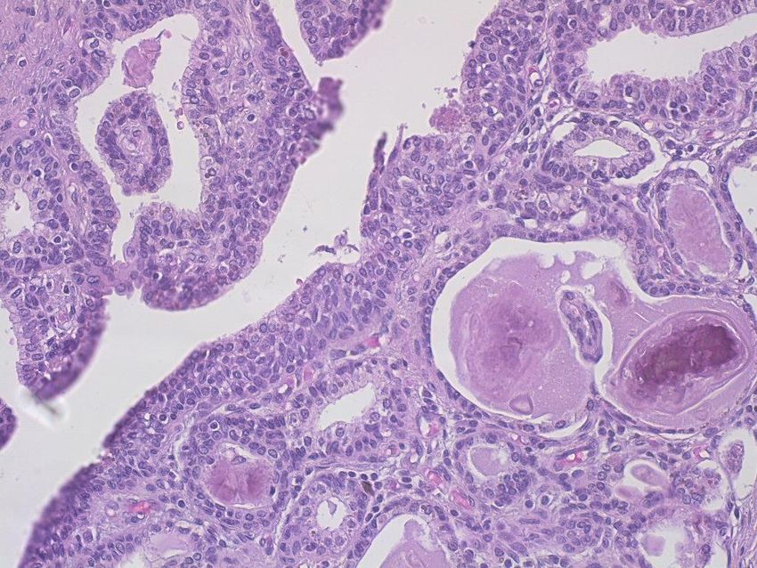
CITOLOGIA URINARIA

  TEST CHE HA L’OBIETTIVO DI
IDENTIFICARE CELLULE ANOMALE
         NELLE URINE

Liquido organico di colore giallastro, maleodorante, a reazione acida,
escreto dal rene a seguito della filtrazione del plasma sanguigno;
è il prodotto finale del processo metabolico; viene espulso dalla
vescica urinaria attraverso l'uretra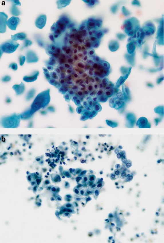
NOSTRO COMPITO

FORNIRE INFORMAZIONI UTILI
/////////////////
                          /////////////////

/////////////////
REFERTO
                                      SICUREZZA
    ?       ?         INFORMAZIONI
                                                   SOGGETTIVITA’

                      COMUNICAZIONE

        ACCURATEZZA                   ESPERIENZA
?                                                        ?
         CHIAREZZA

    ?                 PREDITTIVITA’       ?        FORMAZIONE

                      CONDIVISIONE

?           CREDO                     TRAINING
         CITOLOGICO
                                                     SCUOLA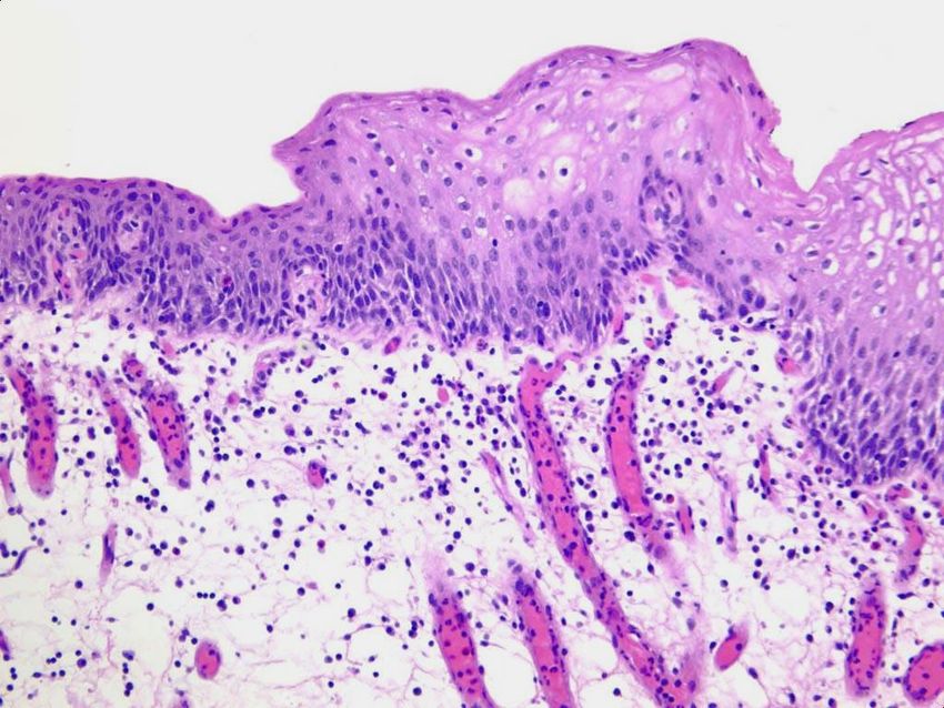
Mirò
Il carnevale di Arlecchino
Anatomia apparato urinario
Citologia normale

CELLULE UROTELIALI   CELLULE GHIANDOLARI

CELLULE SQUAMOSE     CELLULE TUBULARI RENALI
Corredo secondario: granulociti, linfociti, istiociti,
emazie, cristalli, materiale proteico e spermatozoi
CITOLOGIA URINARIA:
     Individuare neoplasie del tratto
                 urinario
a)   Ematuria in pazienti sintomatici
b)   Follow-up
c)   Controllo dopo chemioterapia topica
d)   Professioni a rischio (industrie chimiche e
     metallurgiche...)
Modalità di raccolta
• URINE SPONTANEE
3 campioni di 3 giorni consecutivi
Seconde urine del mattino
Letteratura
>specificità con >nr campioni
• CATETERE
• LAVAGGIO
• NEOVESCICA (vescica ileale)DIFFERENZE MORFOLOGICHE E
METODICA
SPONTANEE CATETERE LAVAGGIO NEOVESCICA
cellularita’ bassa alta piu’ alta alto
preservazione scarsa migliore buona degenerato
architettura cellula singola frammenti gruppi e gruppi e
frammenti singole
tipo cellulare ombrello ombrello, ombrello, enteriche,
basali basali ombrello
vantaggi non invasivo campione ottimo nessuno
migliore
svantaggi degenerazione, artefatti, invasivo degenerazione
contaminazione infezionespontanee catetere lavaggio neovescica
Storia clinica: molto importante!

                Catetere
                Chirurgia
                Calcolosi
                 Terapia

     Per una corretta interpretazione
Allestimento del campione
Varie metodiche:
• Striscio diretto
• Centrifuga e citocentrifuga
• Strato sottile
Colorazione: PapanicolaouLETTURA E REFERTAZIONE
• No atipie citologiche             • Negativo per carcinoma
                                      uroteliale di alto grado
• Modificazioni cellulari benigne   • Cellule uroteliali atipiche
                                    • Sospetto per carcinoma
                                      uroteliale di alto grado
• Atipie indeterminate per
  neoplasia                         • Carcinoma uroteliale di alto
                                      grado
                                    • Neoplasia di basso grado
• Neoplasia di basso grado

                                    • Neoplasie primitive,
• Neoplasia di alto grado             secondarie e miscellanee
ADEGUATEZZA
20 cellule uroteliali ben preservate
X10HPF:
a) 10-20: soddisfacente ma limitato
30 ml… da bassa cellularità
b)Atypical,
ALGORITMO No
(PARIS SYSTEM)
Suspicious,
or
Malignant
Yes
Appropriate
No Instrumented Benign
Urothelial
Yes
Cellularity*
Non–Urothelial
Features
Obscuring Yes
Urothelial
Morphology
Adequate No Yes
No Volume
No
Appropriate
Benign
No
Urothelial
Cellularity*
Inadequate Adequate
Yes YesADEGUATEZZA
(PARIS SYSTEM)
MORFOLOGIA
CELLULARITA’ ADEGUATEZZA VOLUME
RACCOLTAVolume vescica: ml 600
Area superficie interna: 350cm2
Diametro delle cellule uroteliali: micron 200
Urotelio: 5 strati
Numero totale delle cellule è di 10^9:
circa 1.000.000.000
Infinitesima frazione di cellule uroteliali
Basso valore predittivo positivo.
Sensibilità
a)0-73% basso grado
b)95% alto gradoStoria naturale e patogenesi del carcinoma uroteliale
Accrescimento
Papillare Non papillare
Basso grado
I tumori papillari di basso grado
raramente hanno crescita invasiva
Alto grado
I tumori di alto grado hanno crescita invasiva
e possono dare metastasiIl quadro citologico dipende dal pattern di crescita e dal grado di malignità della neoplasia
PARAMETRI CITOLOGICI
cellularità
                     Criterio maggiore:
citoplasma
                     rapporto nucleo-citoplasma
taglia nucleo
                     Criteri minori:
forma nucleo
                     ipercromasia nucleare
nucleoli             membrana nucleare irregolare
                     Cromatina dispersa in modo
cromatina
                     irregolare

N/C

fondo
CATEGORIE DIAGNOSTICHE
Negative for High-Grade
Urothelial Carcinoma
• Cellule benigne uroteliali, squamose e
ghiandolari
• Frammenti di tessuto uroteliale benigno (BUTF)
e aggregati (sheets or clusters)
• Modificazioni associate a litiasi
• Effetto citopatico virale: polyoma virus (BK
virus—decoy cells)
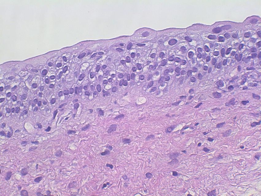
• Effetti post-terapia, inclusa neovescicaFig. 3.2 Intermediate urothelial cells. The intermediate layer of urothelium, immediately underneath the umbrella cells, is easily dissociated into single cells. These often have cells with cytoplasmic (cercariaform) tails (All of the features are normal, and described in Fig. 3.1b .) ( Washing,TP, medium mag. )
Negativo per carcinoma uroteliale di alto grado
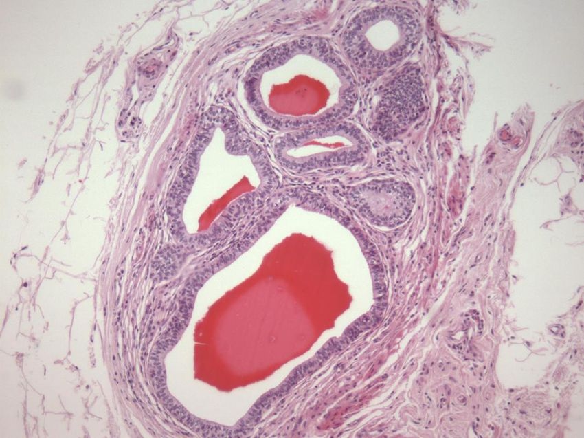
Fig. 3.6 Cystitis cystica/glandularis.
( a ) Glandular cells from the lining of
the bladder can originate from a focus
native to the urothelium, metaplasia
from an inflammatory focus (cystitis
cystica/glandularis), or from a
glandular neoplasm either primary or
secondary. Unless the cytomorphology
suggests a neoplasm, all such TTFs or
cell groups are considered benign
( Washing, TP, high mag. ).
( b ) Cystitis cystica may appear as in
Fig. 3.6 a or as a single layer of
glandular cells. They closely resemble
endocervical cells, and could be from a
case of endocervicosis if the tumor was
also in the muscular wall of the
bladder or ureter. This mucosal strip
was from a focus of cystitis cystica
( Washing, TP, high mag. ).
Cistite ghiandolareNegativo per carcinoma uroteliale di alto
grado
Fig. 3.8 Benign urothelial tissue fragment
(BUTF).
( a ) Voided. BUTF can be seen in voided
urines, and should not mandate a
diagnosis of “atypical”. In this fragment,
nuclei are uniform in
size and shape, evenly spaced, and with
finely granular chromatin ( Voided, SP,
high mag. ).
( b ) Instrumented from renal pelvis. Cell
fragments from the renal pelvis should be
cautiously considered.
In this case, the diagnosis rendered was
“suspicious for low-grade neoplasm”. The
excision of the kidney revealed only
urothelial hyperplasia overlying a
subepithelial hemangioma.
Retrospective review recognized the
uniform nuclear size and round shape.
The resemblance to a papillary lesion was
no doubt the result of instrumentation
( Renal pelvic washing, CS, high mag. )
Benign Urothelial Tissue FragmentsNegativo per carcinoma uroteliale di alto
grado
Fig. 3.10 Urothelium with
nephrolithiasis—threedimensional fragments.
( a ) Kidney and bladder stones can cause
serious changes in the urothelium, sometimes
resembling neoplasms. Careful examination of
the cells in a three-dimensional TTF is critical to
an accurate diagnosis. These cells have round
nuclei which are evenly spaced. Chromatin is
finely granular and nucleoli are inconspicuous.
A renal calculus was discovered on imaging
studies and from the clinical history ( Voided, SP,
moderate mag. ).
( b ) A BUTF in a voided urine may be the result
of numerous causes. In this patient,
nephrolithiasis was the reason. Cellular changes
are mild when compared to those in the photos
to follow (Chap. 4).
The absence of any fibrovascular stalk
eliminates the diagnosis of a low grade LGUN
( Voided, SP, high mag. ).
NefrolitiasiFig. 3.11 Urothelium with nephrolithiasis— sheets or clusters. ( a ) A sheet of urothelium consists of relatively uniform cells with moderately hyperchromatic nuclei. Even though the nuclear chromatin is darker than normal, the presence of a bladder stone is reason enough for the changes. Because of the history and mild changes, this sample was placed in the “Negative” category ( Voided, SP). ( b ) Compare the cells in the center of the field with those to the right, especially considering the nuclear chromatin and nuclear shapes. The central cells are hyperchromatic and the shapes vary. Inflammation is seen in the background. Without the history of nephrolithiasis, these cells would indicate a diagnosis of “atypical urothelial cells” (AUC). If there were any consideration of a urothelial lesion in addition to lithiasis, a note is appropriate or a diagnosis of AUC ( Washing, CS, moderate mag. ).
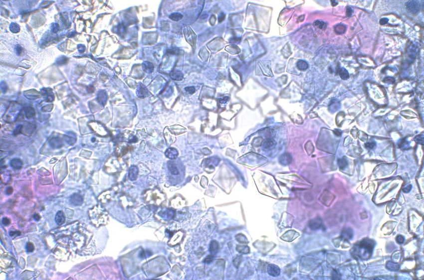
Negative for malignancy. Cells clusters of well-preserved intermediate urothelial cells
have increased N/C ratios. However, hyperchromasia is absent and the nuclei show fine regularly
distributed chromatin with small visible nucleoli. Nuclear membranes are smooth and regular
( Voided urine, TP, high mag. )
RISCHIO DI MALIGNITA’
• Dopo una citologia negativa la biopsia (3.4
  al 6.2 %) rivela una neoplasia di
  basso/alto grado nel 32.2–68.9 %

• Falsi negativi in citologia da neovescica:
  5.7-8.7%
The Paris System for Reporting
                                                      Urinary Cytology
 CATEGORIE DIAGNOSTICHE
 Cellule uroteliali atipiche
Presenza di elementi uroteliali con atipia citologica (non architetturale) di grado
lieve-moderato

CRITERI DIAGNOSTICI

     Maggiore
     Cellule uroteliali non superficiali e non degenerate con un
     aumentato rapporto N/C (>0.5)

     Minori uno richiesto
     Ipercromasia nucleare
     Membrana nucleare irregolare
     Cromatina dispersa in modo irregolare

Note: non include i frammenti senza atipia citologica e nemmeno le cellule uroteliali
sospette per neoplasia
Fig. 4.3 Atypical urothelial cells (AUC). Two groups of urothelial cells are shown. The group at the top is composed of intermediate type urothelial cells with smooth nuclear contours, and no features of atypia. The urothelial cells in the group on the bottom have high N/C ratios, and nuclear contour irregularity. Nuclear chromasia is similar in both groups. Due to the cytologic atypia seen in the group on the bottom this case should be categorized as AUC ( bladder washing, TP, intermediate mag. )
Fig. 4.4 Atypical urothelial cells (AUC). Atypical urothelial cells with high N/C ratios and
nuclear contour irregularity. The absence of hyperchromasia and the presence of degenerated
clumped chromatin preclude a diagnosis of SHGUC ( Bladder washing, TP, high mag. )
Fig. 4.9 Atypical urothelial cells (AUC). Urothelial cells with high N/C ratio (up to 50 %), and
nuclear hyperchromasia are shown. The chromatin is coarse and the nuclear membranes are
irregular. While the features are worrisome for high grade urothelial carcinoma, due to extensive
degeneration and N/C ratio being less than 70 %, AUC diagnosis may be more appropriate.
Follow up showed high grade urothelial carcinoma in the kidney; the urinary bladder had
no pathology. ( Bladder washing, TP, high mag. )
Fig. 4.10 Atypical urothelial cells (AUC). Urothelial cells with high N/C ratios, and nuclear
hyperchromasia. ( c ) Small aggregate of atypical urothelial cells adjacent to squamous cells
(right). The urothelial cell nuclei also show degeneration, but the one cell with the
high N/C ratio is worrisome for carcinoma. The patient is a 36-year-old woman with recurrent
urolithiasis, and no history of urothelial carcinoma. Her age and history are low-risk factors for
bladder cancer. These three fi gures display the entire amount of atypical cells that are present in
the specimen; these cytologic features warrant the diagnosis AUC (Voided,TP, high mag)
RISCHIO CLINICO
• Il rischio di avere una biopsia positiva di
  alto grado dopo una diagnosi di AUC varia
  dall’8.3% al 37.5%
• Inversamente proporzionale al tasso di
  AUC istituzione
• Correlato all’intervallo
• Management basato sul paziente
Sospetto per carcinoma uroteliale
          di alto grado
Presenza di elementi uroteliali non degenerati e non superficiali con le
seguenti caratteristiche citologiche:

criterio diagnostico richiesto
- rapporto nucleo-citoplasma aumentato (0.5-0.7)
- ipercromasia nucleare da moderata a severa

almeno uno dei due seguenti aspetti
- cromatina irregolarmente azzolata
- marcate irregolarità membrana nucleare
Fig. 5.3 Suspicious for high-grade urothelial carcinoma (SHGUC). A few abnormal intermediate urothelial cells, one of which is well preserved (center) and features an increased N/C ratio, hyperchromasia, irregular clumpy chromatin, and severely irregular nuclear membrane. If more than five similar cells were found, the diagnosis of HGUC would be appropriate ( Catheterized urine, CS, high mag. )
Fig. 5.8 Suspicious for high-grade urothelial carcinoma (SHGUC). Rare but abnormal well
preserved intermediate urothelial cells having increased N/C ratios, hyperchromasia, clumpy
chromatin, and irregular nuclear membranes. Note that although the nuclear size is not
significantly larger than normal intermediate cell nuclei, the cells contain cytological nuclear
abnormalities that warrant a “suspicious for high-grade urothelial carcinoma” diagnosis (Voided
urine, SP, high mag)
Fig. 5.4 Suspicious for high-grade urothelial        Fig. 5.10 Atypical urothelial cells (AUC). Cell clusters
carcinoma (SHGUC).                                   of well-preserved intermediate urothelial cells some of
Intermediate urothelial cells showing                which show an increased N/C ratio and hyperchromasia.
increased N/C ratios, hyperchromasia, clumpy         The degree of hyperchromasia is mild in comparison to
chromatin, and irregular nuclear membranes.          the normal intermediate cell nucleus ( upper right ).
Note that not all the cells have an N/C ratio that   In addition, the cells do not show clumpy chromatin
exceeds 0.7 but in the presence of similar nuclear   pattern or irregular nuclear membranes which preclude
characteristics, they should be considered part of    the assignment of a SHGUC diagnosis ( Voided urine,
the same lesion. A “positive for HGUC” diagnosis     TP, high mag. )
may be acceptable in this case,especially in the
presence of a previous history of HGUC
(Catheterized urine, CS, high mag. )
Fig. 5.9 Suspicious for high-grade urothelial     Fig. 5.11 Atypical urothelial cells (AUC).
carcinoma (SHGUC). Rare but abnormal well         Abnormal intermediate urothelial cells showing increased
Preserved intermediate urothelial cells showing   N/C ratio and irregular nuclear membranes in the
increased N/C ratios, hyperchromasia, irregular   absence of nuclear hyperchromasia preclude a diagnosis
Nuclear membranes but overall fine evenly         of SHGUC. The follow-up diagnosis was LGUC
distributed chromatin                             ( Voided urine, TP, high mag. )
(Voided urine, TP, medium mag)
RISCHIO DI MALIGNITA’

• Il rischio di avere una biopsia positiva per
  neoplasia di alto grado dopo una diagnosi di
  sospetto varia dal 37.8% al 95%

• Il valore predittivo positivo: 79% (sotto i sei mesi)
  e 80% intorno ai 6 mesi) contro l’86% e il 90% del
  positivo per alto grado
High-Grade Urothelial
        Carcinoma (HGUC)

a) Cellularità: 5-10
b) Rapporto nucleo-citoplasma elevato (0.7)
c) Ipercromasia nucleare moderata-severa
d) Membrana nucleare irregolare
e) Cromatina irregolare (grossolana/azzollata)
f) Aspetti secondari:pleomorfismo, dismetrie,
necrosi, mitosi, ecc……
Fig. 6.2 High-grade urothelial carcinoma (HGUC) present as a cohesive group of malignant cells. The N/C ratio of 0.7 is noted in the majority of the tumor cells ( Bladder washing, TP, high mag. )
Fig. 6.3 Nuclear hyperchromasia is present in this cell from a patient with high-grade
urothelialcarcinoma (HGUC). Note the tumor necrosis clinging to the cells ( Bladder washing,
TP, high mag. )
Fig. 6.9 A few cells exhibit classic features of high-grade urothelial carcinoma (HGUC)
adjacent to cells of squamous differentiation ( Bladder washing, TP, high mag. )
Fig. 6.11 High-grade urothelial carcinoma (HGUC) tumor cells with glandular differentiation
Are from the same sample as Fig. 6.11 ( Bladder washing, TP, high mag. )
RISCHIO CLINICO
“cystoscopic examination with biopsies”
Neoplasia uroteliale di basso grado

      Aggregati tridimensionali di cellule uroteliali (papille)
    comprensive di asse fibrovascolare che include i capillari
Fig. 7.1 Positive for LGUN (composite). ( a ) Highly cellular specimen composed of numerous tissue fragments. ( b )–( d ) Some fragments show three-dimensional papillary configuration. Fibrovascular cores are appreciated in the center of papillary structures ( Renal pelvic washing, CS, ( a )–( c ) low mag. ( d ) medium mag. )
Fig. 7.2 Positive for LGUN. Three-dimensional papillary structures have central cores. Notice
mild cytologic atypia and disorganization of cells forming papillae. Photo courtesy of David
Wilbur ( Renal pelvic washing, CS, medium mag. )
Fig. 7.3 Positive for LGUN.
( a ) Three-dimensional cluster
of cells with nuclear overlapping, forming
papillae. There is a thin capillary vessel running
through the center of the cluster ( Washing, TP,
low mag .). ( b ) Positive for LGUN. Occasionally,
 if there is enough material left in a container,
 a cell block may be helpful to visualize
fibrovascular cores
( Washing, Cell block, H&E stain, low mag. )
Fig. 7.4 Negative for HGUC with a comment suggestive of LGUN. Ill-defined three-dimensional
papillary structure may represent a LGUN. No obvious capillary vessel is seen. Accumulation of
red blood cells in the middle of the cluster resembles the outline of the blood vessel wall
( Washing, TP, medium mag. )
PROGRESSIONE
• Papilloma: 0%

• Neoplasia papillare uroteliale a basso
  potenziale:3.6%

• Carcinoma uroteliale di basso grado: 5-
  25%
DISTRIBUZIONE CATEGORIE
Total range    Academic Private practice
Positive (%)     1.0–6.3         1.2–4.9
Suspicious (%)   0.7–5.4         0.2–2.7
Atypical (%)     1.8–23.7      3.1–21.4
Negative (%)    75.4–94.8     71.4–96.1
Washing (%)      1.0–74        1.0–22.2
Number/year 841–9210          81–4932
SISTEMA DI PARIGI
• Standardizzazione linguaggio

• Malattie clinicamente rilevanti (alto grado)

• Definizione di rischio per singola categoria

• Riduzione categorie borderline
GRAZIE
Puoi anche leggere