Giustificazione, ottimizzazione e livelli diagnostici di
       riferimento alla luce del D.Lgs. 101/2020
←
→
Trascrizione del contenuto della pagina
Se il tuo browser non visualizza correttamente la pagina, ti preghiamo di leggere il contenuto della pagina quaggiù
Giustificazione, ottimizzazione e livelli diagnostici di
riferimento alla luce del D.Lgs. 101/2020
The Simpsons’ Guide to Radiation
Radiation exposure and risk

 Stochastic risk:
    has probability of occurrence depending on the irradiated doses
    without threshold
    shows up years after exposure
    consists primarily of cancer and genetic effects such as inherited
    mutations
 Deterministic effects:
    malfunctions of organs by irradiation more than a threshold
    skin burns, cataract, cardiovascular diseases, intestinal damage,
    hemopoietic system and CNS system failure
Source of ionizing radiation to the
population

Radiation exposure and risk

                 100 mSv
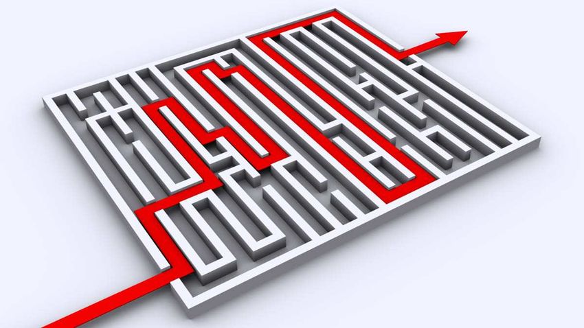
Risk estimation: why so difficult?

 Extremely large sample size needed to ensure statistical
 significance at low dose levels:
    sample size of 500.000 and 2.000.000 are required with lifetime
    follow-up for exposure levels of 20 mSv and 10 mSv respectively,
    which rend a decent epidemiological study unfeasible*
 Uncertainties in radiation dosimetry cannot be avoidable
 and influence every aspect of studies
 Issues of confounding factors such as smoking, genetic
 variation and socioeconomic status are very important
 Statistical uncertainties in dose response model
          *National Research Council (US) Committee on Assessment of CDC Radiation Studies, 1995
Risk estimation: why so difficult?

 Despite a variety of studies, understanding of health
 effects of low dose radiation – less than 100 mSv – is
 still incomplete
 For this reason, the LNT approach is the most
 reasonable risk model at low dose levels and remain
 fundamental in terms of radiation protection and safety

D.Lgs. 101/2020. Art.1 – Finalità e
principi del sistema di radioprotezione

 Il sistema di radioprotezione si basa sui principi di
 giustificazione, ottimizzazione e limitazione delle dosi
 …le esposizioni mediche non sono soggette a limitazioni
 delle dosi…
Justification

 Any exposure from diagnostic imaging is justified if it can
 provide the benefits of a prompt diagnosis and adequate
 treatment: these benefits always outweigh any
 associated risk such as a small additional risk of cancer
 due to the exposure to radiation
D.Lgs 101/2020 - Art. 4: Giustificazione delle
pratiche

 1. Nuovi tipi di pratiche … debbono essere giustificate
 prima di essere adottate
 2. Le pratiche esistenti sono sottoposte a riesame ogni
 qualvolta emergano nuove evidenze sulla loro efficacia e
 potenziali conseguenze, ovvero si rendano disponibili
 altre pratiche …
Art. 156 e 157 – Ambito di applicazione
del principio di giustificazione

 pazienti nell’ambito della rispettiva diagnosi o
 trattamento
 sorveglianza sanitaria dei lavoratori
 persone nell’ambito di screening sanitari
 asintomatici e pazienti che volontariamente partecipano
 a programmi di ricerca
 persone nell’ambito di procedure a scopo non medico
 condotte con attrezzature radiologiche
Art. 156 e 157 – Ambito di applicazione
del principio di giustificazione

 E’ vietata l’esposizione non giustificata
 Tutte le esposizioni mediche individuali devono essere
 giustificate preliminarmente, tenendo conto degli
 obbiettivi specifici dell’esposizione e delle caratteristiche
 della persona interessata. Una pratica non giustificata in
 generale potrebbe esserlo nel singolo individuo in
 circostanze particolari
Art. 161 – Procedure

 Il Ministero della salute… adotta linee guida per le
 procedure inerenti le pratiche radiologiche clinicamente
 sperimentate e standardizzate
 Nelle linee guida sono altresì fornite raccomandazioni ai
 medici prescriventi relative ai criteri di appropriatezza e
 giustificazione, nonchè indicazioni sull’entità delle dosi
 assorbite dai pazienti... Tali linee guida sono pubblicate
 nella Gazzetta Ufficiale
 Fino alla pubblicazione in GU…
Adapted from
EC RP 118, 2000
SIRM, AINR, AIMN, FISM, SIMI,
ISS, Ministero della Salute, ASSR

                          (2004)
ESR iGuide is based on the Appropriateness Criteria developed by the American College of Radiology (ACR), reviewed by a team of senior radiologists Recommendations for topic groups including Breast, Cardiac, Gastrointestinal, Musculoskeletal, Neurologic, Paediatric, Thoracic, Urologic, Vascular and Women’s Imaging are provided Separate guidance for children includes 320 indications/scenarios with a number of 2465 scored decision rules Annual update are provided in cooperation with the ACR’s Rapid Response Committee
•   1800 indications with associated exams including appropriateness
    ratings for defined patient groups
     – Age range 0-150 years
     – Sex: male female, unknown
•   Age range 0-18: paediatric guidelines
     – 320 indications, 2465 scored rules
•   Appropriateness ratings:
     – 1-3 (red): usually not appropriate
     – 4-6 (yellow): may be appropriate
     – 7-9 (green) usually appropriate
ESR iGuide workflow
ESR iGuide: an example of clinical scenario.
First febrile urinary infection in a 6-y-old male

                                                    Relative Radiation
                                Clinical scenario          Level
              Appropriateness

                                Imaging study
ESR iGuide: head trauma in a 1-y-old male
ESR iGuide: chronic abdominal pain in a 7-y-old male
ESR iGuide implementation
Croatia pilot project (ECR 2019)
             Appropriateness of referrals with ESR iGuide
           November 2016-May 2018
          Approximately 100.000 decision support sessions for all modalities
Art. 166 – Protezione particolare durante la gravidanza e l’allattamento • In gravidanza il medico specialista porrà particolare attenzione alla giustificazione, alla necessità o all’urgenza, considerando la possibilità di procrastinare l’indagine. Nel caso in cui l’indagine diagnostica non possa essere procrastinata, il medico specialista informa la donna dei rischi derivanti al nascituro. Nel caso in cui si debba procedere comunque all’esposizione, il medico specialista e il tecnico sanitario di radiologia medica devono porre particolare attenzione al processo di ottimizzazione riguardante sia la madre che il nascituro
Optimisation

 All doses due to medical exposure for radiodiagnostic,
 interventional radiology,…, are kept As Low As
 Reasonably Achievable consistent with obtaining the
 required medical information, taking into account
 economic and societal factors

                    ALARA
Optimisation
240 mAs 120 mAs
Optimisation means applying the ALARA concept
Applying the ALARA concept means using a sound
technique and accepting the highest image noise consistent
with obtaining the required medical informationArt. 158 – Applicazione del principio di ottimizzazione alle esposizioni mediche Tutte le dosi dovute alle esposizioni di cui all’articolo 156, a eccezione delle procedure radioterapeutiche, devono essere mantenute al livello più basso ragionevolmente ottenibile e compatibile con il raggiungimento dell’informazione diagnostica richiesta, tenendo conto di fattori economici e sociali
Art. 158 – Applicazione del principio di
ottimizzazione alle esposizioni mediche

Il responsabile dell’impianto radiologico, ai fini
dell’ottimizzazione dell’esecuzione degli esami in radio-
diagnostica… nonchè delle procedure di radiologia
interventistica, garantisce che si tenga conto dei livelli
diagnostici di riferimento, laddove disponibili, tenendo
conto delle indicazioni più aggiornate pubblicate
dall’Istituto Superiore di Sanità
Art. 165 – Pratiche speciali

L’esercente e il responsabile dell’impianto radiologico,
nell’ambito delle rispettive competenze, individuano
gli interventi da attuarsi ai fini dell’applicazione del
principio di giustificazione e di ottimizzazione alle
pratiche che comportano, in particolare, esposizioni
di soggetti:
a) in età pediatrica;
b) esposti nell’ambito di programmi di screening;
c) esposti nell’ambito di pratiche radiologiche comportanti alte
dosi quali: radiologia interventistica, TC, medicina nucleare;
d) sottoposti a trattamenti radioterapeutici
Livelli diagnostici di riferimento (LDR)

Introdotti nel 1996 nella pubblicazione ICRP n 73
Definiti come (Art. 7 D.Lgs 101/2020) come “livelli di
dose nelle pratiche radiodiagnostiche mediche o
interventistiche… per esami tipici per gruppi di pazienti di
corporatura standard o fantocci standard”
NON SI APPLICANO AL SINGOLO PAZIENTE E
NON RAPPRESENTANO LIMITI DI DOSE
Livelli diagnostici di riferimento (LDR)

Rappresentano uno strumento essenziale nei
processi di ottimizzazione delle esposizioni,
individuando quelle pratiche radiologiche che richiedono
interventi tecnici e/o metodologici atti a ridurre la dose
mediana ai pazienti sottoposti ad indagine diagnostica in
una installazione radiologica
Qualora il valore di LDR venga ecceduto in modo
significativo deve essere intrapresa una revisione e
vengono adottate azioni correttive (Art. 161 D.Lgs
101/2020)
Responsabilità del RIR e dello specialista in fisica medica
Procedure di interesse per gli LDR (ISTISAN 20/22)

 Gli LDR vengono determinati per esami che:
   sono eseguiti spesso (almeno 15 pazienti in un bimestre
   all’interno della struttura)
   hanno una denominazione univoca
   consentono di eseguire verifiche in una elevata percentuale di
   installazioni radiologiche
   o erogano una dose potenzialmente elevata
Gli LDR nella pratica clinica (ISTISAN 20/22)
ISTISAN 20/22
ISTISAN 20/22
Existing DRLs set in
children by competent
 authorities for body
regions for CT studies

•   DRLs set by an authoritative
    body

•   Other published/available data

•   Not available

    Radiation Protection N° 185, 2018
• Despite recommendations, few paediatric DRLs are set in less than half of EU countries, and many of them are obsolete • Paediatric DRLs should have been implemented by February 2018 (BSS Euratom directive 2013/59)
• Why?
 – the number of paediatric examinations is lower than
   in adults
 – the paucity of dose data in children makes difficult to
   collect sufficient data to establish DRLs
DRLs: grouping in children

 Traditionally based on age
 Children can vary in weight
 by a factor of 200:
   premature baby (400 gr)
   obese adolescent (>80 kg)
European Commission RP 185 (2018)

http://www.eurosafeimaging.org/wp/wp-content/uploads/2018/09/rp_185.pdf
European DRLs for radiography and
fluoroscopy in children (RP 185)
European DRLs for radiography and
fluoroscopy in children (RP 185)
European Diagnostic Reference Levels

• European DRLs: based
  on the median (the 50th
  percentile) value of the
  distribution of the NDRLs
  for a defined clinical
  imaging task surveyed for
  standardised patient
  groupings
• 16 cm phantom for head
  studies, 32 cm phantom
  for chest and abdomen
  studies
• These values refer to a
  single acquisition, not to
  the entire examination
ISTISAN 20/22
ISTISAN 20/22
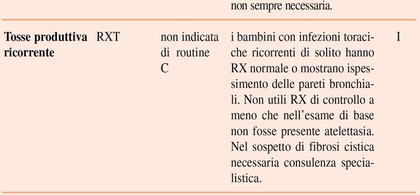
Gli LDR nella pratica clinica (ISTISAN 20/22)

Un LDR è da considerarsi superato quando il valore
mediano per un campione rappresentativo di
pazienti di corporatura normale, oppure di pazienti
all’interno di un intervallo eventualmente specificato di
peso e/o dimensioni e/o età, è maggiore del
corrispondente valore di LDR
La propria pratica radiologica deve essere confrontata
con gli LDR disponibili almeno ogni 4 anni per la
radiologia convenzionale, ogni due anni per la TC e
annualmente per la radiologia interventistica
Gli LDR nella pratica clinica (Art. 164 e allegato
XXVIII D.Lgs 101/2020)

Nel manuale di qualità della struttura vanno inseriti i
riferimenti bibliografici dai quali sono stati tratti gli LDR e
i risultati della verifica degli LDR, da conservare almeno
10 anni
DRLs: present limitations

                            Hydrocephalus
    Head trauma with
   epidural haematoma
DRLs: present limitations
Kidney stones HaepatoblastomaDRLs: present limitations • Presently, DRLs for CT studies are established in relation to body region • It is common experience that in “real life” CT protocols are differentiated – and consequently delivered dose – according to the clinical indication of the study • Clinical based DLRs for CT studies are presently missing • EC funded EUCLID European Study on Clinical DRLs in adults presently ongoing • A similar study in children is very much needed
Radiation protection mainstays

                                 DRLs
Radiation Risk and children
Children are potentially more vulnerable to radiation
exposure:
they grow quickly, and their cells are more sensitive to radiation
have longer lifespans to develop long-term radiation-induced
detrimental effects
are more vulnerable than adults to the development of certain
cancer typesRadiation Risk
Radiation Risk
Radiation Risk
Euratom Directive 2013/59/EURATOM:
Justification

Article 55 requires that Medical exposure shall show a
sufficient net benefit, weighing the total potential
diagnostic or therapeutic benefits it produces… against
the individual detriment that the exposure might cause,
taking into account... alternative techniques having the
same objective but involving no or less exposure to
ionising radiation
Article 57 requires that the referrer and the practitioner
are involved, as specified by Member States, in the
justification process of individual medical exposures
Euratom Directive 2013/59/EURATOM:
Referral Guidelines

Article 58 requires that Member States shall ensure
that referral guidelines for medical imaging, taking into
account the radiation doses, are available to the
referrers
Referral guidelines for diagnostic
imaging

 Referral guidelines for diagnostic imaging support the
 best use of clinical radiology as long as they:
    conform to the best evidence-based standards
    protects the patient from unnecessary exposure to ionising
    radiation
    provide dedicated guidance for children and pregnant
    women/unborn child
    provide the evidence for which imaging resources can be
    used efficiently and effectively
Radiation protection N°178 (2014) RP 178 (2014) provides information on Referral Guidelines for Medical Imaging availability and use in the European Union based on a European-wide survey
Availability of RG in Europe

 30 European Countries provided information

              5

                                          Guidelines available
                                          Guidelines not available
                            25

                                              RP 178, 2014
Availability of separate guidance for
children
Only 12 out of 30 EU Countries provided information
8
6
4
2
0
Available
Not available
Don't know
RP 178, 2014Availability of separate guidance for
pregnant women /unborn children
• Only 12 answers were provided
8
7
6
5
4
3
2
1
0
Available
Not available
RP 178, 2014Guidelines availability and update
It appears from RP 178 document that in several
European countries referral guidelines for
children may be not available or regularly
updated
The year of the first edition of imaging guidelines varied
from 1989 to 2005
The approximate duration of the review cycle has varied
between countries from 3-4 years to > 6 years, being in
some countries older than 10 years
RP 178, 2014RP 178: conclusions and recommendations • Imaging referral guidelines were available in most European countries, although in many cases detailed information about these guidelines were not provided • A single set of European guidelines should be preferred • National guidelines, developed de novo through accepted methodology or adopted or adapted are alternatives • Separate advice for children and pregnant women / unborn child must be included • Additional measures are needed to reinforce the use of guidelines • Clinical Decision Support systems interfacing with RIS and electronic requesting systems should be implemented
ESR iGuide is freely available through the ESR website
since 2018
ESR iGuide is the ESR solution to make imaging referral
guidelines – including separate guidance for children
– readily available and easily usable across Europe
ESR iGuide guidelines are embedded in a clinical decision
support platform, which allows users to localise the
recommendations according to their needs starting from an
evidence-based-core
ESR iGuide is designed to be a user-friendly system
available to referring physicians at the point of care
Euratom Directive 2013/59/EURATOM:
Optimisation

Article 56 requires that all doses due to medical
exposure for radiodiagnostic, interventional radiology,…,
are kept As Low As Reasonably Achievable consistent
with obtaining the required medical information, taking
into account economic and societal factors

                   ALARA
Euratom Directive 2013/59/EURATOM: Diagnostic Reference Levels (DRLs) Article 56 requires that Member States shall ensure the establishment, regular review and use of DRLs for radiodiagnostic examinations, having regard to the recommended European DRLs where available… National DRLs: dose levels in diagnostic practices for typical examinations for groups of standard-sized patients or standard phantoms for broadly defined types of equipment. These levels are expected not to be exceeded for standard procedures when good and normal practice regarding diagnostic and technical performance is applied
Setting of DRLs

• National DRLs (NDRLs):
  – based on the 3rd quartile value of the median values
    of the distributions of patient doses from a
    representative sample of RX departments in the
    country, for a defined clinical imaging task surveyed
    for standardised patient groupings
  – set by an authoritative body, based on national
    patient surveys
  – NDRLs should be compared with the European DRLs
  – institutions must carry out regular comparison of their
    LDRLs with NDRLs
Setting of DRLs
• Local DRLs (LDRLs):
• based on the median values of patient dose
distribution from examinations from the healthcare
facility
• set by a given hospital or group of hospitals for their
own use to improve optimisation
• set to correspond to the level of technology and local
achievements of optimisationSetting of DRLs
• European DRLs (EDRLs):
– based on the median value of the distribution of
NDRLs for a defined clinical imaging task surveyed for
standardised patient groupings
– EDRLs provide an interim solution for countries with
no NDRLs, until such NDRLs become availableDosimetric quantities to be used • Radiography: – PKA, (Ka,e) • Fluoroscopy and IR: – PKA, (Ka,r, fluoroscopy time, number of images) • CT: – CTDIvol referred to phantom size (16 or 32 cm) – DLP
Recommended patient grouping

• Chest and abdomen:
  – weight
  – age (to be used just to make comparison between old and
    new paediatric DRLs)
• Head:
  – age
ISTISAN 17/33
ISTISAN 17/33
DRLs and Dose Management Systems

DMSs allow automatic recording, retrieval and analysis of
dosimetric data from radiological studies
They allow establishing of local DRLs, which can be used
for comparison with national or European DRLs
DMSs are an excellent tool for optimisation and
compliance with established DRLs
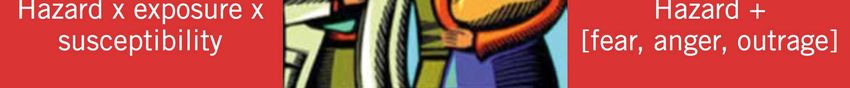
“Harm and alarm”

 • “… about 1500 of those children will die later in
   life from radiation induced cancer…”
                                   USA Today, January 22nd, 2001
Communicating risk to parents
WHO, 2016Responsibilities
• Directive 2013/59/EURATOM:
– Art. 56 (1 d): wherever practicable and prior to the
exposure taking place, the practitioner or the referee,
as specified by Member States, ensures that the
patient or their representatives is provided with
adequate information relating to the benefits and
risks associated with the radiation dose from the
medical exposure. Similar information as well as
relevant guidance shall be given to carers and
comforters…Establishing a patient-centred communication Speak slowly, use plain language and avoid medical terms Explain the rationale of the procedure, emphasizing its benefits Illustrate the potential risks by comparing them with other kinds of common risks Explain what will be done to minimize risk to the patient Repeat key messages Encourage questions, and be prepared to address them Cards/leaflets for patients/parents may be helpful
Some practical examples

Questions                                  Possible response
Why is this radiological examination       This examination can rapidly clarify
recommended?                               your child’s diagnosis
Is there any risk from this radiological   One concern is the possibility of cancer
Questions
examination                                Possible response
                                           resulting  from the radiation
How great is this risk?                    The  risk is
                                           The risk  ofvery
                                                         missing   a serious
                                                              small,          diagnosis
                                                                      if any. We   are not
                                           will
                                           sureoccur    now. The
                                                 that there         potential
                                                              is a risk  at veryeffects
                                                                                 low of
                                           radiation
                                           dose, like –those
                                                         if anywith
                                                                 - would
                                                                     CT ortake
                                                                             most X-ray
                                           years/decades
                                           studies
When will these risks occur?               The risk of missing a serious diagnosis
                                           will occur now. The potential effects of
                                           radiation – if any - would take
                                           years/decades
Some practical examples
Questions Possible response
Why can’t we do a procedure that We have considered using
does not use radiation instead? examinations that do not require
radiation, but we have determined this
is the best procedure to answer the
Questions clinical
Possiblequestion
responseand plan treatment
Can
Doesthe
mydose
childbe adjusted
need so that
it? Does s/he my There
need The are many
referring techniques
practitioner to lower
and
child receives the lowest possible
it now? dose and risk
radiologist without
have done acompromising
risk-benefit
dose? the diagnostic
analysis quality
and this of images.
specific procedureOuris
facility uses appropriate
recommended protocolsand
to aid in diagnosis for
children
treatment
What are the consequences of not Your child’s health may be affected
doing the procedure? through incorrect or delayed diagnosis
and treatment
Adapted from Broder et al, 2014, and WHO, 2016Some practical examples
Typical Effective
Pediatric examination Risk
Dose (mSv)
XR Arm, Dental, Skull, Chest Negligible hours/days Chance of dying
< 0.02
from flu,
firework
3 years 10 x less than lifetime risk
>10
of dying in motor
vehicle accident
> 500 CXRSome practical examples
• A 2-year-old child underwent CT scan of the skull,
chest and abdomen after an accident. The family
doctor stated the following:
• a) the CT scan has possibly tripled the risk for your
child of developing cancer within 18 years of age
(from 0.5% to 1.5%)
• b) the CT scan proved essential to evaluate your
child’s conditions and to treat his wounds, which
otherwise would have put his health at risk. The
probability of your child having a normal
development has remained almost the sameResources
http://www.eurosafeimaging.orgTake home points Understanding of health effects of low dose radiation – as used in medical imaging – is still incomplete For this reason, the linear no-threshold NT approach is the most reasonable risk model at low dose levels Justification and optimisation are the mainstays of radiation protection DRLs and dose management systems are excellent tools for optimisation Effective communication of benefits and risk of medical imaging to parents is fundamental
Puoi anche leggere