ITALIAN JOURNAL OF EMERGENCY MEDICINE - itjem
←
→
Trascrizione del contenuto della pagina
Se il tuo browser non visualizza correttamente la pagina, ti preghiamo di leggere il contenuto della pagina quaggiù
Anno IV, numero 1 - Febbraio 2015

        ITALIAN JOURNAL OF
        EMERGENCY MEDICINE
Official Journal of the Italian Society of Emergency Medicine

    3    Notizie dall’ufficio stampa

    4    Sulle tracce dell’ECG

    8    Special Articles

   16    Articoli originali

   42    Area Giovani

   45    Letteratura in Urgenza
ITALIAN JOURNAL OF
        EMERGENCY MEDICINE
                                www.itjem.org - e-mail: info@itjem.org

                       Official Journal of Italian Society of Emergency Medicine
Registrazione del Tribunale di Milano, 401 24/06/2008 - Ownership and Copyrigth - Electronic Edition

       SIMEU, Società Italiana di Medicina di Emergenza-Urgenza, Via Vittor Pisani 10, Milano

                 The Electronic Edition is available at www.cgems.it e www.simeu.it
Article disseminated via www.cgems.it are abstracted, indexed and referensed by many abstracting
       and information Service biblography networks, subscription agencies, library networks

                           EDITOR IN CHIEF                SCIENTIFIC BOARD
                           Cinzia Barletta                Giuliano Bertazzoni
                                                          Maria Antonietta Bressan
                           SCIENTIFIC MANAGER             Giorgio Carbone
                           Paolo Groff                    Gianalfonso Cibinel
                                                          Andrea Dellepiane
                           EDITORIAL BOARD                Annamaria Ferrari
                           Andrea Fabbri                  Nicola Filippo Glorioso
                           Paolo Balzaretti               Paola Noto
                           Roberto Cosentini              Franco Perraro
                           Fabio De Iaco                  Riccardo Pini
                           Mauro Fallani                  Alessandra Revello
                           Rodolfo Ferrari                Adelina Ricciardelli
                           Luca Gelati                    Gianfranco Sanson
                           Federica Stella                Fernando Schiraldi
                           Sossio Serra
                           Isabella Di Zio                PRESS OFFICE
                                                          Silvia Alparone

             PUBLISHING MANAGER                            PUBLISHER
             Gianpiero Garnero                             CG Edizioni medico Scientifiche srl
             CG. Edizioni Medico Scientifiche srl          Via Piedicavallo, 14 - 10145 Torino
             Via Piedicavallo, 14 - 10145 Torino           Tel. 011.33.85.07 r.a.
             Tel. 011.33.85.07 r.a. - Fax 011.38.52.750    Fax 011.38.52.750
             E-mail: garnero@cgems.it                      Web: www.cgems.it - www.cgems.eu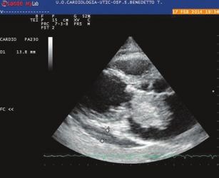
Sommario
3 Notizie dall’ufficio stampa
Una partnership con Cittadinanzattiva per la Settimana nazionale Simeu del Pronto Soccorso, 16-24 maggio 2015
Silvia Alparone
4 Sulle tracce dell’ECG
Sulle tracce dell’ECG: Torsade de pointes
Mauro Fallani, Isabella Di Zio, Sossio Serra, Federica Stella
8 Special Article
Bowel obstruction: the role of MSCT in emergency department
Emanuela Capalbo, Farideh Sajadidehkordi, Anna Kluzer, Paola Mariani, Maurizio Cariati
16 Articoli originali
Arresto cardiocircolatorio nel territorio di modena e provincia: uno studio retrospettivo osservazionale
Brugioni Lucio, Gozzi Cristina, Serantoni Carlo, Silvestri Alessandra, Casini Francesco, Loschi Giuseppe
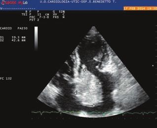
Identificazione di una massa intracardiaca con ecocardiografia “fast” eseguita in fase di triage da uno studente di
infermieristica all’interno del Dipartimento di Emergenza. Caso Clinico
Vito Maurizio Parato, Patricia Ciapanna, Gloria D’Angelo, Rita Elia, Noemi Minichelli, Erica Ciccarelli, Mariella
Amadio, Tiziana Traini, Valentina Simonetti, Silvano Troiani, Paolo Groff
Linee di indirizzo per la gestione della Colite Ulcerosa severa nel DEA: il tempo è colon?
Marina Rizzi, Francesco Panzera, Carmine Sinno
Management del paziente con pericardite acuta in pronto soccorso:
metodologia di approccio in area critica e successivo iter clinico-assistenziale
a cura di Pasquale De Luca
con la collaborazione di: Antonio De Luca, Vito Sollazzo, Antonio Manfrini, Stefano Carughi,
Marco Sperandeo, Riccardo Ieva, Gianluigi Vendemiale, Tommaso Lancialonga
42 Area giovani
Trombosi acuta della vena mesenterica superiore: case report
L. Pagani, S. Paiardi, A. Raimondi, S. Marra, G. Ricevuti, M.A. Bressan
45 Letteratura in Urgenza
Noninvasive Ventilation and Survival in Acute Care Settings: A Comprehensive Systematic Reveiew and Meta-
Analysis of Randomized Controlled Trials. Luca Cabrini, et al. Critical Care Medicine 2015; in press
Top 10 ideas to improve your bedside teaching in a busy emergency department. Gary M Green, Esther H Chen.
Emergency Medicine Journal 2015;32:76
A Clinical Classification of the Acute Respiratory Distress Syndrome for Predicting Outcome and Guiding Medical
Therapy. Jesùs Villar, et al, for the Acute Lung Injury: Epidemiology and Natural history (AILEN) Network. Critical
Care Medicine 2015;43:346
The use of non-invasive ventilation in very old patients with hypercapnic acute respiratory failure because of COPD
exacerbation. A Nicolini, et al. The International Journal of Clinical Practice 2014 Dec;60(12):1523 doi: 10.1111/
ijcp.12484
Commento a cura di Rodolfo FerrariNotizie dall’ufficio stampa 3
Una partnership con Cittadinanzattiva per la Settimana nazionale Simeu
del Pronto Soccorso, 16-24 maggio 2015
Silvia Alparone
Giornalista
Torna anche quest’anno la Settimana nazionale Simeu del Pronto Soccorso: dal 16 al 24 maggio 2015 sul territo-
rio di tutte le regioni italiane, la Società Italiana di Medicina di Emergenza-Urgenza organizzerà incontri e attività
per favorire il confronto fra i professionisti sanitari dell’emergenza e la popolazione, in un’occasione lontana dalla
stringente necessità dei momenti di urgenza sanitaria.
Come già per la Settimana dello scorso anno, i luoghi scelti saranno piazze, teatri, centri commerciali, scuole, cen-
tri sportivi: non quindi gli ospedali e gli spazi tradizionali per l’attività sanitaria, ma gli spazi della cittadinanza.
La novità principale dell’edizione di quest’anno è la collaborazione con Cittadinanzattiva – Tribunale dei diritti
del Malato: se la Settimana del Pronto soccorso 2014 ha sottolineato l’importanza di un patto di alleanza fra
cittadini e professionisti sanitari dell’emergenza, l’edizione 2015 dà sostanza a quel patto, con una partnership
ufficiale fra Simeu e la principale associazione nazionale dei pazienti. Il Tribunale dei diritti del Malato, iniziativa
di Cittadinanzattiva, insignito della Medaglia d’oro al merito della sanità pubblica dal Presidente della Repub-
blica nel 2006, conta in Italia 21 sedi regionali e circa 300 assemblee locali. Ha come obiettivo “di contribuire
a una più umana, efficace e razionale organizzazione del servizio sanitario nazionale, coinvolgendo cittadini e
operatori dei servizi”. La rete del Tribunale, come rete di tutela, si integra con la rete di cura, costituita dai 331
Dea e 513 Pronto Soccorso distribuiti sul territorio nazionale. L’alleanza tra le due associazioni, entrambe attente
alla centralità del paziente nel percorso di cura in emergenza, ma anche all’importanza dell’operatore sanitario,
parte attiva nello stesso percorso e a sua volta cittadino, ha come obiettivo la creazione di una squadra, forte e
tenace, che possa dal confronto fare proposte concrete condivise di miglioramento e cura dell’organizzazione del
PS, con ricadute positive su cittadini, operatori sanitari-cittadini, quindi sull’intera comunità.
La partnership fra Simeu e TdM in occasione della Settimana del Pronto Soccorso si concretizzerà nel lancio di un
primo progetto condiviso, un monitoraggio Simeu-Cittadinanza Attiva: in molte strutture di emergenza nazionali
verranno distribuite due schede, preparate in collaborazione fra le due associazioni, su due aspetti critici per
l’emergenza-urgenza nazionale, il servizio offerto al cittadino in Pronto Soccorso e l’organizzazione dei Pronto
Soccorso.
I temi identificati come concetti guida della manifestazione Simeu di quest’anno sono la centralità del Pronto Soc-
corso all’interno del Servizio Sanitario Nazionale, come estremo baluardo di un servizio pubblico sempre più in
difficoltà, e la gestione del dolore in emergenza.
Il gruppo nazionale Simeu che lavora all’organizzazione dell’edizione 2015 della Settimana PS è composto da
Paola Caporaletti, Mario Guarino, Paolo Cremonesi, Antonella Cocorocchio, Giuseppe Pepe, dalla segreteria e
da Silvia Alparone. Coordina Maria Pia Ruggieri, segretario nazionale della Società scientifica.
Per ulteriori informazioni: info@simeu.it.
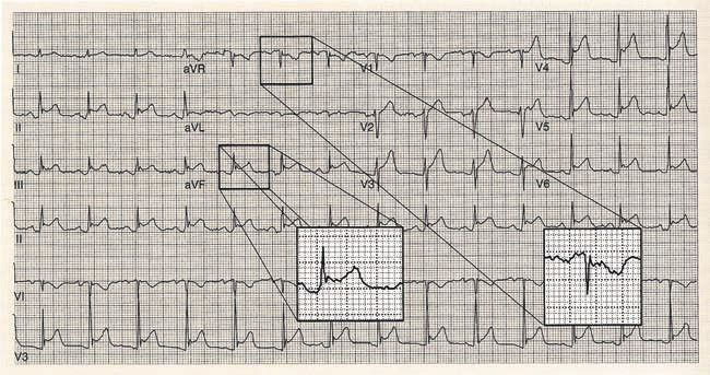
ITALIAN JOURNAL OF EMERGENCY MEDICINE - Febbraio 20154 Sulle tracce dell’ECG Sulle tracce dell’ECG: Torsade de pointes Mauro Fallani*, Isabella Di Zio**, Sossio Serra***, Federica Stella**** * Responsabile UOS Medicina d’urgenza, Ospedale “Ceccarini” di Riccione, AUSL Rimini ** Dirigente Medico Medicina d’urgenza, Pronto Soccorso Ospedale “Madonna del Soccorso” di San Benedetto del Tronto *** Dirigente Medico Medicina d’urgenza, Pronto Soccorso, Ospedale “M. Bufalini” di Cesena **** Medico in Formazione Specialistica, Scuola di Specializzazione in Medicina di Emergenza-Urgenza, Università degli Studi di Padova Parole chiave: torsione di punta, sindromi del QT lungo, allungamento del QT. Keywords: Torsades de pointes, Long QT Syndrome, QT-prolongation. Caso Clinico Una paziente di 76 anni, con storia di cardiopatia ischemica ed evoluzione dilatativa (FE 33%) e portatrice di pacemaker-PM per episodi di blocco atrio-ventricolare (BAV) avanzato viene ricoverata in Medicina d’Urgenza per episodio sincopale in corso di iperpiressia. La prima ipotesi diagnostica è che la perdita di conoscenza sia le- gata all’iperpiressia anche perché il PM è stato controllato 2 settimane prima. La paziente da 2 giorni è in terapia antibiotica con amoxicillina-acido clavulanico 1 gr x 3 ed azitromicina 500 per il riscontro di un focolaio bronco pneumonico; assume inoltre furosemide 75 mg die, ramipril 5 mg die, bisoprololo 5 mg die. L’esame obbiettivo polmonare supporta la diagnosi posta per la presenza di crepitazioni alla base polmonare destra e gli esami ese- guiti documentano una leucocitosi neutrofila (GB 12.500 con neutrofili 86%) una normale funzionalità epatica e renale e una lieve ipokaliemia (3.2 mEq/l). La paziente viene collegata alla telemetria ECG e praticamente imme- diatamente presenta nuovo episodio sincopale di breve durata; il riesame della monitorizzazione ECG documenta una tachicardia ventricolare polimorfa con la continuo mutare dell’ampiezza e dell’asse del QRS, compatibile con una torsade de pointes (Fig. 1). La torsade ed il QT lungo La “torsades de pointes” è una tachicardia ventricolare polimorfa, spesso sostenuta da bradicardia ed associata ad allungamento del QT, che prende nome dal periodico cambiamento di ampiezza e polarità dei complessi QRS intorno alla linea isoelettrica, con il vettore del QRS da muta da positivo a negativo e viceversa, e che nell’insieme ricorda un nastro che si torce. Di solito l’aritmia tende ad autolimitarsi spontaneamente, peraltro con tendenza a recidivare, ma può anche degenerare in fibrillazione ventricolare. La sua presentazione può essere quella della morte improvvisa in paziente con cuore strutturalmente sano; per questo è ovvio che la sua prevenzione ha una importanza strategica [1-2]. Morfologicamente si presenta come una tachicardia a complessi larghi con morfologia del QRS diversa da battito a battito ed una frequenza compresa fra 150 e 250 battiti al minuto, spesso con un allungamento del QT all’ECG nella fase non critica. Se da tempo era nota l’associazione fra QT lungo e torsade i meccanismi coinvolti sono diventati chiari solo negli ultimi anni. Fondamentalmente sono delle alterazioni dei canali ionici che provocano durante la ripolarizzazione un rallentamento dei flussi ionici; tale situazione induce dei potenziali di azione precoci, denominati early after depolarizations (EAD). Il rallentamento della ripolarizzazione, causa poi degli EAD, non si verifica in tutto il mio- cardio, essendo l’endocardio profondo e lo strato intermedio dei ventricoli maggiormente proni: la differenza fra zone in cui si realizzano gli EAD e il resto del miocardio provoca dei fenomeni di rientro che sono il trigger per l’innesco della torsade. Fra l’altro il non omogeneo rallentamento della ripolarizzazione in differenti zone del miocardio provoca il fe- nomeno della dispersione del QT per il quale la misurazione del QT è diversa nelle singole derivazione; inoltre maggiore è la dispersione più è facile l’insorgenza della torsade. Sono al momento note almeno 8 varianti genetiche associate al QT lungo con alterazione dei canali ionici e ten- ITALIAN JOURNAL OF EMERGENCY MEDICINE - Febbraio 2015
Sulle tracce dell’ECG                  5

Figura 1. Torsione di punta, registrazione al monitor.

denza ad innescare la torsade; tali condizioni sono classificate come canalopatie assieme alla sindrome di Bruga-
da, il QT corto e la tachicardia ventricolare polimorma catecolaminergica [3]. Ritornando alle varianti di QT lungo
nei genotipi LQT1, LQT2, LQT5, LQT6 e LQT7 l’alterazione è a livello dei canali del potassio, mentre nella variante
LQT3 il difetto è a livello dei canali del sodio e nelle varianti LQT4 e LQT8 i canali alterati sono quelli del calcio.
Dal punto di vista epidemiologico è interessante notare come l’intervallo QT corretto è maggiore nella popolazio-
ne bianca rispetto alla popolazione nera, e più lungo nelle femmine rispetto ai maschi. Pertanto, la torsades de
pointes è più comune nella razza bianca e nelle femmine. Da segnalare poi come la torsades si verifica a qualsiasi
età; se si verifica in età precoce, la causa è di solito dovuta alla sindrome del QT lungo congenita. Negli anni
successivi, la causa è di solito a causa di sindrome del QT lungo acquisita.
E’ ovvio come a questo punto sia necessaria una breve digressione sul QT e sulla sua misurazione: l’intervallo QT
è il periodo compreso fra l’inizio del QRS e la fine dell’onda T. La sua misurazione è resa ardua sia dalla disper-
sione del QT, già descritta e dalla difficoltà a stabilire la fine della onda T, specie in presenza di onda U; onda U
è evidente in alcuni soggetti senza patologia oppure in presenza di ipokaliemia (situazione a sua volta associata
ad allungamento del QT).

                                                                              ITALIAN JOURNAL OF EMERGENCY MEDICINE - Febbraio 2015
6 Sulle tracce dell’ECG
La normale durata del QT è influenzata dalla frequenza cardiaca tanto che è necessario calcolare il QT corretto
per la frequenza (QTc) in base alla formula secodo la quale QTc = QT/radice quadrata RR (ove sia il QT che
l’RR sono espressi in secondi). Solo per la frequenza di 60 bpm il QT coincide con il QTc (poiché l’RR espresso
in secondi è 1 e così pure la sua radice quadrata). Per le altre frequenze il calcolo è più complesso tanto che, se
pur non metodologicamente corretto, si fa riferimento a valori normali del QT in funzione della frequenza come
nella tab. 1.
Tab. 1.
frequenza bpm QT (msec)
50 400
60 380
70 360
80 340
90 320
100 300
110 290
120 280
130 260
Ad ogni buon conto nel sospetto di allungamento del QT è essenziale calcolare il QTc e verificarne la patologicità
per sesso ed età con la tab. 2.
Tab. 2.
Età 1-15 Maschi adulti Femmine adulte
normale < 440 msec < 430 msec < 450 msec
borderline 440-460 msec 430-450 msec 450-470 msec
patologico > 460 msec > 450 msec > 470 msec
Le cause di QT lungo e conseguentemente torsade possono essere classificate come congenite (la sindrome di
Jervell e Lange-Nielsen, autosomica recessiva, associata a sordità e la sindrome di Romano-Ward, autosomica
dominante senza sordità) oppure acquisite; fra queste ultime le più comuni sono legate all’uso di farmaci che
allungano il QT (4-5), il cui elenco è consultabile al indirizzo www.qtdrugs.org e alterazioni elettrolitiche quali
l’ipokaliemia e l’ipomagnesiemia.
Terapia
Bisogna dividere la gestione in acuto da quella in cronico. Nella gestione del fatto acuto se l’aritmia non si auto-
limita sono necessarie le manovre rianimatorie con eventuale defibrillazione in caso di degenerazione in fibrilla-
zione ventricolare. Nelle fase intercritiche la somministrazione di solfato di magnesio alla dose di 1-2 gr per via
endovenosa in 30-60 sec (ripetibile dopo 5-15 min) riduce l’inducibilità della aritmia, sopprimendo i potenziali
che innescano l’aritmia (opzione efficace anche nel caso descritto, assieme alla somministrazione di potassio e la
sospensione del macrolide). I pazienti trattati vanno accuratamente seguiti per il rischio di depressione neuromu-
scolare indotta dal magnesio. Anche il mantenimento di un corretto valore di K+ plasmatico ha un effetto analogo.
Nel caso di refrattarietà alle terapie descritte con reiterazione delle fasi di tachicardia ventricolare è indicato,
nelle forme acquisite, l’aumento della frequenza cardiaca; questa può essere realizzata, nelle forme acquisite,
utilizzando farmaci adrenergici (l’isoprotenerolo per esempio). I farmaci adrenergici aumentano la conduzione
ITALIAN JOURNAL OF EMERGENCY MEDICINE - Febbraio 2015Sulle tracce dell’ECG 7
atrio-ventricolare, e aumentando la frequenza cardiaca riducono il QT e la dispersione della durata della ripola-
rizzazione. Nelle forme di QT lungo congenito la frequenza cardiaca deve essere ottenuta con il pacing (anche
transcutaneo nelle emergenze). Il pacing è utilizzabile anche nelle forme di QT lungo congenito; l’efficacia del
pacing è legata all’aumento della frequenza cardiaca che riduce il QT. Il pacing atriale è la modalita di scelta,
mantenendo il contributo atriale al riempimento ventricolare ed il QRS stretto.
Nelle forme congenite la terapia b-bloccante alla massima dose tollerabile è indicata con propanololo o nadololo
a meno di severa bradicardia preesistente.
Sia nelle forme congenite che in quelle acquisite ove si renda indispensabile la somministrazione di farmaci poten-
zialmente scatenanti è indicata la valutazione per l’impianto di un defibrillatore impiantabile.
Bibliografia
Dave J, Bessette MJ, Setnik G, et al. Torsade de Pointes. http://emedicine.medscape.com/article/1950863-overview
Eleftherios M, Kallergis, Christos A, Goudis, Emmanuel N, Simantirakis, et al. Mechanisms, Risk Factors, and Manage-
ment of Acquired Long QT Syndrome: A Comprehensive Review. The Scientific World Journal Volume 2012, Article
ID 212178, 8 pages doi: 10.1100/2012/212178
Roberts JD, Gollob MH. The genetic and clinical features of cardiac channelopathies. Future Cardiol. 2010 Jul; 6(4):
491-506. doi: 10.2217/fca.10.27.
Roden DM. Drug-induced prolongation of the QT interval. NEJM 2004; 350: 1013-1022.
Fenichel RR. Drug-induced Torsades de pointes and implication for drug development. J Cardiovascular Electroph 2004;
15: 475-495.
ITALIAN JOURNAL OF EMERGENCY MEDICINE - Febbraio 20158 Special Articles Bowel obstruction: the role of MSCT in emergency department Emanuela Capalbo, Farideh Sajadidehkordi, Anna Kluzer, Paola Mariani, Maurizio Cariati Scuola di Specializzazione di Radiodiagnostica, Università degli Studi di Milano. Ospedale San Carlo Borromeo via Pio II 3, Milano 20153 Italy Abstract Objectives: evaluating diagnostic performance of multi-slice computed tomography (MSCT) in emergency de- partment (ED), in characterization of site and type of bowel obstruction and comparing with surgical findings. Methods: we selected 119 patients positive for bowel obstruction. The surgical treatment is performed up to 48 hours after imaging. We obtained the written informed consent by patients and Ethical Committee approval. We calculated sensitivity, specificity, positive (PPV) and negative (NPV) predictive value and diagnostic accuracy. Dia- gnostic concordance of cause and site has been evaluated using K Cohen’s coefficient. Results: the main causes of bowel obstruction, according to MSCT are: adhesion 21%, inflammation 13,4%, tumors 10,9%, volvulus-intussusception 10,9%. MSCT for diagnosis of cause has a sensitivity of 96,9%, specificity 85%, PPV 96,9%, NPV 85% and diagnostic accuracy 94,9%.The localization was in duodenum 15,9%, jejunum 12,6%, ileum 17,6%, right colon 15,1%, left colon 13,4% and sigma/rectum 9,2%. MSCT for diagnosis of site has a sensitivity of 97,5%, specificity 87,2%, PPV 97,8%, NPV 86,5% and diagnostic accuracy 96,1%.The cause and site is indeterminate in 15,9%. K=0,75 for cause and 0,71 for site of bowel obstruction. Conclusions: MSCT identifies with a good concordance both cause and localization of the bowel obstruction compared with surgical diagnosis. It is diagnostic reference-standard in ED to define the patient’s therapy planning. Keywords: MSCT, diagnosis, emergency, bowel occlusion. Introduzione Since the introduction of multi-slice computed tomography (MSCT), a progressive increase in the use of this method has been registered for both elective and emergency evaluation of intestinal pathology, and in particular where there is suspicion of bowel obstruction. Many emergency-urgency conditions occur with acute abdominal pain, among them there is the bowel obstruction that occurs also with vomiting and bowel closed by feces and gas [1]. In many cases it is difficult to make a dia- gnosis based only on the clinical and laboratory tests; to this end the imaging has often a decisive role [2]. Radiological exams used in these cases are: X-ray, ultrasound and MSCT of the abdomen, and the choice between them depends on the severity of symptoms, on the anamnesis of patient and on the decision of the clinician and the radiologist. The MSCT provides a lot of information in short scan times and allows evaluating the site of mechanical occlusion and whether it is or not associated with intestinal ischemia. This will help in the choice between medical or surgical treatment, and in the latter case between laparotomy or laparoscopy [3]. Bowel obstruction may be localized at small or large intestine and, according to the cause; it is classified in mecha- nical (adhesions, internal and external hernias, diverticulitis, tumors, volvulus, gallstone ileus, endometriosis and chronic inflammatory diseases) and dynamic. The dynamic ileus is the result of a significant intestinal contraction [1] or the evolution of a mechanical ileus unrecognized or not promptly treated [4]. Adhesions and hernias are the most common causes of small bowel obstruction, reported by approximately 80% of cases [5]. Tumors, sigmoid diverticulitis and volvulus are the most common causes of large bowel obstruction and together constitute 80-85% of cases [6]. Aim of this paper is to evaluate the diagnostic performance of MSCT carried out in the emergency department (ED) in making an accurate diagnosis of cause and location of intestinal obstruction, correlating the radiological diagnosis with surgical findings. Italian Journal of Emergency Medicine - Febbraio 2015
Special Articles               9

Material and methods
Selection of Patients
In 2013 we recorded 8256 patients in emergency Radiology for non-traumatic acute abdominal pain; 1372 of
these performed a MSCT (16,7%).
For this study, we selected retrospectively 119 patients with bowel obstruction. We included patients with high
suspicious of bowel occlusion at triage, and/or after surgery visit and radiological diagnosis. Then we examined
the medical records considering the surgery as reference diagnostic. The surgical treatment has been performed up
to 48 hours after imaging. We obtained the written informed consent by patients and Ethical Committee approval
for this research study.
MSCTs have been evaluated by many radiologists, with years of experience ranging from 5 to 20, during different
work shifts at ED. All medical reports described the location and cause of bowel obstruction. Subsequently, the
images were reviewed in blind by two experienced radiologists (18 and 20 years of experience) who pointed out
the presence of signs in the MSCT, suggestive of bowel obstruction.
For the radiological diagnosis of obstruction cause, we grouped the lesions as follows: cancer, inflammation
(mainly diverticulitis and appendicitis), adhesions, internal and external hernias, volvulus and intussusception,
gallstone ileus, endometriosis, inflammatory bowel disease (Crohn’s disease and ulcerative colitis), fitobezoar/
tricobezoar, and external bodies.
The site of occlusion was divided into: duodenum, jejunum, ileum, right colon (from cecum to transverse colon
included), left colon (from splenic flexure to the sigmoid excluded) and rectum/sigma.

TC method
The examinations were performed using a 16-slice CT Lightespeed (GE Medical System, Milwauke WI, USA). The
protocol foresees an acquisition from the diaphragmatic dome to the pelvis in basal conditions and, based on the
clinical suspicion and according to the indications given in literature [7], intravenously administration of non-ionic
iodinated contrast medium (concentration 370 mg/dl, dosage 1,5-2ml/kg, flow 2,5-3ml/sec). In such cases after
injection of contrast medium we completed the exam with an early acquisition after 35 sec (arterial phase) and/or
late acquisition after 90 seconds (portal phase).
We set the following parameters: collimation 16x0.75mm, FOV 320-360mm, pitch 1,75, voltage 120kV, auto-
matic modulation of tube current (mA), slice thickness 3,75 mm, imaging reconstruction to 1,25 or 2,5mm with
a standard filter for multiplanar evaluations, display window for abdomen. In no case the contrast medium was
administered orally or rectally.

Statistical Analysis
The data were analyzed using SPSS version 18.
The baseline characteristics of the study population were expressed as mean±standard deviation for continuous
variables, and as percentages for categorical variables. The diagnostic concordance between radiological and
surgical diagnosis was evaluated by using the K test of Cohen, both for the cause and the site of the occlusion.
The concordance was also calculated for the retrospective diagnosis performed by radiologists A and B, as well
as between the original and the retrospective diagnosis. Finally, we calculated sensitivity, specificity, diagnostic
accuracy, positive (PPV) and negative (NPV) predictive value.

Results
The general characteristics of the patients included in this study are summarized in Tab. 1.
In 97 patients out of the 102, the cause of the occlusion described by MSCT was confirmed by surgical diagnosis
(Fig. 1-2). In 3 patients there was a misdiagnosis of endometriosis, adherence and inflammation which were not
seen in the intervention.
In 2 patients we had an incorrect diagnosis of the cause: an inflammation with abscess and an external body were
found to be respectively a tumor and a gallstone ileus in surgery.

                                                                             Italian Journal of Emergency Medicine - Febbraio 2015
10 Special Articles
Tabella 1. General characteristics of the patiens included in the study.
Patients 119
M/F (%) 67/52 (56,3/43,7)
Mean Age(±DS) 68,7 (±23,8)
Surgical 89 (74,7)
Triage (%)
Medical 30 (25,3)
Surgical treatment (%) 102 (85,7)
MSTC with contrast ev (%) 97 (81,5)
A B C
Figura 1. MSCT scans of an occlusive condition. !a: axial acquisition; 1b,1c: MPR acquisitions.
A B C
Figura 2. MSCT scan of an occlusive condition. 2a,2b: axial acquisitions;
2c: MPR acquisition.
In 19 patients included in the study, MSCT has not provided a precise diagnosis of neither site nor the cause of the
occlusion. In 17 of these patients, surgeons decided to perform optic colonoscopy. Since the result was negative,
they behaved in a “wait and see” manner: 7 were sub-occlusive forms which resolved spontaneously and 10 were
inflammation which resolved with appropriate medical therapy.
Italian Journal of Emergency Medicine - Febbraio 2015Special Articles 11
Tabella 2. Concordance between MSCT and surgery per diagnosis.
MSCT (%) SURGERY (%)
Tumors 13 (10,9) 15 (14,7)
Inflammation 16 (13,4) 14 (13,7)
Adhesion 25 (21) 24+1* (23,5)
Hernias 9 (7,5) 9 (7,5)
Volvulus and Intussusception 13 (10,9) 13 (12,7)
Gallstone Ileus 5 (4,2) 6 (5,2)
Endometriosis 3 (2,5) 2 (1,9)
Inflammatory Bowel Disease 7 (5,8) 7 (6,8)
Fitobezoar and Tricobezoar 4 (3,3) 5 (4,9)
Exsternal body 5 (4,2) 4 (3,9)
Indetefinite/Negative 19 (15,9) 3 (2,9)
TOTAL 119 (100) 102+1*(100)
*diagnosis made only by surgery
Tabella 3. Concordance between MSCT and surgery per site.
MSTC (%) SURGERY (%)
Duodenum 19 (15,9) 19 (18,6)
Jejunum 15 (12,6) 16 (15,6)
Ileum 21 (17,6) 20 (19,6)
Right Colon 18 (15,1) 19 (18,6)
Left Colon 16 (13,4) 16 (15,6)
Sigma/Rectum 11 (9,2) 12 (11,7)
Indeterminate/Negative 19 (15,9) 3 (2,9)
TOTAL 119 (100) 102 (100)
In the remaining 2 patients, the persistence and severity of the symptoms guided the surgeons to perform an explo-
rative laparoscopy, identifying an occlusive disease: it was one case of fitobezoar located in the cecum in a young
patient and one tumor in the rectum in a woman with diverticular disease.
The diagnosis of cause performed with MSCT has a sensitivity of 97%, with a specificity of 77,2%, PPV and NPV
of 95% and 85% respectively, with a diagnostic accuracy of 95,7%. The concordance between MSCT and surgery
has a k=0,75 and p12 Special Articles
Tabella 4. Concordance between radiologist A and radiologist B as to the presence of major obsturction signs.
Radiologist A (%) Radiologist B (%)
Bowel Dilatation (>2,5 cm SB, > 6 cm LB) 56 (47) 58 (48,7)
Transition Zone 47 (39,5) 50 (42)
Bowel Feces sign 21 (17,6) 19 (15,9)
Whirlpool sign 11 (9,2) 8 (6,7)
Pneumatosis 8 (6,7) 8 (6,7)
Thickening wall 19 (15,9) 18 (15,1)
Low/absent enhancement wall 21 (17,6) 20 (18,8)
Mesenterial vessels congestion 9 (7,5) 8 (6,7)
Effusion intra-abdominal 28 (23,5) 28 (23,5)
Tabella 5. Inter-radiologist concordance per cause and site.
RADIOLOGIST A (%) RADIOLOGIST B (%)
Cause Site Cause Site
SENSIBILITY 98,1 97,5 97,9 97,8
SPECIFICITY 78,3 87,2 78,5 87,2
PPV 95,7 97,8 95,5 97,6
NPV 86,2 86,5 86,3 86,5
DIAGNOSTIC ACCURACY 97 96,1 97,2 96
Tabella 6. Comparison of single acquisitions vs. both assesements in the diagnosis of cause and site.
AXIAL MPR (cor+sag) AXIAL+MPR
Cause Side Cause Side Cause Side
SENSIBILITY 96 95,3 95,8 96 97 96.9
SPECIFICITY 75,6 84 76 84,6 77,2 85
PPV 94,7 95,1 94 94,8 95 96,9
NPV 83,9 84,3 84,6 84 85 85
DIAGNOSTIC ACCURACY 95,3 93,7 94,9 93,1 95,7 94,9
The concordance between original diagnosis (performed by more radiologists during multiple work shifts in ED)
and retrospective diagnosis (performed by two radiologists, A and B) of cause and site were found to have a k-
value of 0,77 and 0,80 respectively.
In addition, we calculated sensitivity, specificity, PPV, NPV and diagnostic accuracy, considering only axial acquisi-
tions (Fig. 1a, 2a, 2b) or only the MPR (Fig. 1b, 1c, 2c): in both cases, the evaluation of only one of the two modes
has provided results slightly lower than the results of both assessments together (pSpecial Articles 13
Tabella 7. Types of vascular occlusion.
VASCULAR OCCLUSION N (%)
Mesenteric artery 9 (47,4)
Celiac origin 3 (15,8)
Portal Vein 4 (21,1)
Mesenteric vein 2 (10,5)
Mesenteric vein+splenic vein 1 (5,2)
Total 19 (100%)
Acquisitions after contrast medium diagnosed the presence of intestinal ischemia in 16,8% of the study population,
which was confirmed in 100% of cases at surgery intervention. The vascular occlusions are reported in Tab. 7.
Discussion
Intestinal occlusion is frequent in emergency practice, with incidence of the hospitalizations for acute abdomen
of 20% [8, 9]. It is a significant cause of morbidity and mortality and an appropriate treatment depends by early
diagnosis and by accurate identification of patients who require surgery [5].
In literature the MSCT is described able to exclude the disease or identify site, cause and severity [9, 10] and it
is useful in the therapeutic decision-making [11, 12] as it helps the surgeon to determine the type of intervention
[13]. It is a highly accurate method in the diagnosis of occlusion, with a sensitivity, specificity and accuracy of
respectively 90-96%, 96% and 95% [14, 10].
In our study, MSCT (Fig. 1, 2) performed in patients with clinical suspicion of intestinal obstruction confirmed high
sensitivity, specificity and diagnostic accuracy (Tab. 6).
The diagnosis of occlusion by MSCT is based on the identification of dilated proximally intestinal loops and col-
lapsed distally intestinal loops; identifying the point where there is change of intestinal caliber is critical (Fig. 1, 2)
[15, 11]. The interpretation of axial images is essential (Fig. 1a, 2a, 2b), although the associated analysis of the
MPR (Fig. 1b, 1c, 2c) helps in recognizing the obstruction localization [16, 17, 12,13].
The two modalities improve the visualization of the transition zone (Fig. 1c) [15, 18, 19, 20, 11, 14, 15, 16]
with accuracy between 90 [20, 16] and 94% [15, 11], in agreement with values obtained in our study (Tab. 6).
According to Filippone [21, 17], small intestine occlusions (Fig. 2c) are better diagnosed by axial acquisitions
than the coronal reconstructions; however large intestine occlusions (Fig.1c) are better diagnosed by coronal re-
constructions.
Among the various radiological signs of intestinal obstruction the “sign of the feces” (Fig. 1C) (presence of structu-
red material within dilated loop of small intestine, immediately upstream of the transition zone) has been described
as little sensible and specific for the diagnosis of occlusion especially of the small intestine [21, 22, 23, 17, 18,
19], in fact it can be present even in the absence of occlusion [15, 11]. While according to Wang [13] it would
be predictive of intestinal ischemia [13, 2,3 20, 19]. In our study, we found it in 7 of 19 patients (36,8%) with
ischemia (Tab. 1). Pneumatosis has been described as predictive sign of ischemia, but its absence does not exclude
the presence of ischemia [6]. Among 8 patients with pneumatosis described in our study, 5 had ischemia (Tab. 1).
The “whirlpool sign”, originally defined by Fisher as “loop of the small intestine coiled around superior mesenteric
artery” [24, 21], was also associated with volvulus of the large intestine [25, 22]. According to literature, diagno-
stic value for volvulus of “whirlpool sign” is limited: we found it only in 23% of patients with volvulus (Tab. 1, 3).
The intra-abdominal effusion is a sign of a serious occlusive condition and it is often associated with ischemia [6].
80% of patients with intra-abdominal effusion had ischemia; the other 20% had cancer or inflammation. In several
studies the accuracy of MSCT was difficult to assess objectively because of the variable elapse between the radio-
logical examination and surgical intervention [13, 20]. This bias is not present in our study because the maximum
time between the MSCT and the surgery is 48 hours.
Italian Journal of Emergency Medicine - Febbraio 201514 Special Articles
In this study we found a good concordance between the retrospective and the initial diagnosis, according to Wang
[13, 20]. A good knowhow of the TC signs and TC characteristics of the bowel obstruction [26, 23] is essential for
a correct diagnosis and appropriate treatment [3].
Limitations
The limitations of this paper is the relatively small number of patients analysed and the possible selection bias due
to the retrospective study.
Conclusions
MSTC performed in emergency has a high diagnostic accuracy in diagnosis of cause and site of intestinal obstruc-
tion, reducing morbidity and mortality.
We showed a high concordance between radiological and surgical diagnosis, also thanks to the use of the MPR.
Therefore, nowadays the MSCT represents the reference-standard for the diagnosis of intestinal obstruction.
References
1. Harrison TR (2005) Principi di Medicina Interna.Vol II, 16rd edn, McGraw-Hill Companies, srl Publishing Italia,
Milano.
2. Unni K, Udayasankar Jianhai Li DA, Baumgarten WC et al. Acute abdominal pain: value of non contrast enhanced
ultra-low-dose multi detector row CT as a substitute for abdominal radiographs. Emerg Radiol 2009; 16: 61-70.
3. Hayakawa K, Tanikake M, Yoshida S et al. Radiological diagnosis of large-bowel obstruction: non neoplastic etiol-
ogy. Jpn J Radiol 2012; 30: 541-552.
4. Passariello R, Simonetti G. Compendio di Radiologia, 3rd edn, Idelson-Gnocchi,Napoli 2010.
5. Santillan CS. Computed Tomography of Small Bowel Obstruction. Radiol Clin N Am 2013; 51: 17-27.
6 aourel P, Kessler N, Lesnik A et al. Helical CT of large bowel obstruction. Abdom Imaging 2001; 28: 267-75.
7. Leschka S, Alkadhi H, Wildermuth S et al. Multi-detector computed tomography of acute abdomen. Eur Radio 2005;
15: 2435-2447.
8. Scaglione M, Grassi R, Pinto A et al. Positive predictive value and negative predictive value of spiral CT in the diag-
nosis of closed loop obstruction complicated by intestinal ischemia. Radiol Med 2004; 107 (1-2): 69-77.
9. Nicolaou S, Kai B, Ho S et al Imaging of acute small-bowel obstruction. AJR Am J Roentgenol 2005; 185 (4): 1036-
44.
10. Qalbani A, Paushter D, Dachman AH. Multidetector Row CT of Small Bowel Obstruction. Radiol Clin North Am.
2007; 45 (3): 499-512.
11. Rosen MP, Sands DZ, 3rd et al. Impact of abdominal CT on the management of patients presenting to the emergency
department with acute abdominal pain. AJR Am J Roentgenol 2000; 174: 1391-1396.
12. Udayasankar UK, Li J, Baumgarten DA et al. Acute abdominal pain: value of non-contrast enhanced ultra-low-dose
multi-detector row CT as a substitute for abdominal radiographs. Emerg Radiol 2009; 16: 61-70.
13. Wang Q, Chavhan GB, Babyn PS et al. Utility of CT in the diagnosis and management of small-bowel obstruction
in children. Pediatr Radiol. 2012; 42: 1441-1448.
14. Silva AC, Pimenta M, Guimaraes LS. Small bowel obstruction: what to look for. Radiographics. 2009; 29: 423–439
15. Angelelli G, Moschetta M, Cosmo T et al. CT Diagnosis of the nature of bowel obstruction: morphological evaluation
of the transition point. Radiol Med. 2012; 117: 749-758.
16. Horton KM, Fishman EK. The current status of multidetector row CT and three-dimensional imaging of the small bowel.
Radiol Clin North Am. 2003; 41 (2): 199-212.
17. Jaffe TA, Martin LC, Thomas J et al. Small-bowel obstruction: coronal reformations from isotropic voxels at 16-section
multi-detector row CT. Radiology. 2006; 238 (1): 135-42.
18. Aufort S, Charra L, Lesnik A et al. Multidetector CT of bowel obstruction: value of post-processing. Eur Radiol. 2005;
15: 2323-2329.
19. Sinha R, Verma R. Multidetector row computed tomography in bowel obstruction. Part 1. Small-bowel obstruction.
Clin Radiol. 2005; 60: 1058-1067.
Italian Journal of Emergency Medicine - Febbraio 2015Special Articles 15
20. Hodel J, Zins M, Desmottes L et al. Location of the transition zone in CT of of bowel obstruction: added value of
multiplanar reformations. Abdon Imaging. 2009; 34: 35-41.
21. Filippone A, Canci R, Storto ML. Bowel obstruction: comparison between multidetector-row Ct axial and coronal
planes. Abdon Imaging. 2007; 32-310-316.
22. Catalano O. The feces sign: a Ct findings in small bowel obstruction. Radiology. 1997; 37: 417-419.
23. Lazarus DE, Slywotsky C, BBennett GL et al. Frequency and relevance of the “small-bowel feces” sign on CT in pa-
tients with small bowel-obstruction. AJR. 2004; 183: 1361-1366.
24. Fisher JK. Computed tomographic diagnosis of volvulus in intestinal malrotation. Radiology. 1991; 140: 145-146.
25. Gollub MJ, Sora Y, Smith L McG et al. Does the CT whirl sign really predict small bowel volvulus? Experience in an
oncologic population. J Comput Assist Tomogr. 2006; 30: 25-32.
26. Hayakawa K, Tanikake M, Yoshida S et al. Radiological diagnosis of large-bowel obstruction: non neoplastic etiol-
ogy. Jpn J Radiol. 2012; 30: 541-52.
Italian Journal of Emergency Medicine - Febbraio 20158 Special Articles Bowel obstruction: the role of MSCT in emergency department Dr. Emanuela Capalbo Scuola di Specializzazione di Radiodiagnostica, Università degli Studi di Milano. Ospedale San Carlo Borromeo via Pio II 3, Milano 20153 Italy Abstract Objectives: evaluating diagnostic performance of multi-slice computed tomography (MSCT) in emergency de- partment (ED), in characterization of site and type of bowel obstruction and comparing with surgical findings. Methods: we selected 119 patients positive for bowel obstruction. The surgical treatment is performed up to 48 hours after imaging. We obtained the written informed consent by patients and Ethical Committee approval. We calculated sensitivity, specificity, positive (PPV) and negative (NPV) predictive value and diagnostic accuracy. Dia- gnostic concordance of cause and site has been evaluated using K Cohen’s coefficient. Results: the main causes of bowel obstruction, according to MSCT are: adhesion 21%, inflammation 13,4%, tumors 10,9%, volvulus-intussusception 10,9%. MSCT for diagnosis of cause has a sensitivity of 96,9%, specificity 85%, PPV 96,9%, NPV 85% and diagnostic accuracy 94,9%.The localization was in duodenum 15,9%, jejunum 12,6%, ileum 17,6%, right colon 15,1%, left colon 13,4% and sigma/rectum 9,2%. MSCT for diagnosis of site has a sensitivity of 97,5%, specificity 87,2%, PPV 97,8%, NPV 86,5% and diagnostic accuracy 96,1%.The cause and site is indeterminate in 15,9%. K=0,75 for cause and 0,71 for site of bowel obstruction. Conclusions: MSCT identifies with a good concordance both cause and localization of the bowel obstruction compared with surgical diagnosis. It is diagnostic reference-standard in ED to define the patient’s therapy planning. Keywords: MSCT, diagnosis, emergency, bowel occlusion. Introduzione Since the introduction of multi-slice computed tomography (MSCT), a progressive increase in the use of this method has been registered for both elective and emergency evaluation of intestinal pathology, and in particular where there is suspicion of bowel obstruction. Many emergency-urgency conditions occur with acute abdominal pain, among them there is the bowel obstruction that occurs also with vomiting and bowel closed by feces and gas [1]. In many cases it is difficult to make a dia- gnosis based only on the clinical and laboratory tests; to this end the imaging has often a decisive role [2]. Radiological exams used in these cases are: X-ray, ultrasound and MSCT of the abdomen, and the choice between them depends on the severity of symptoms, on the anamnesis of patient and on the decision of the clinician and the radiologist. The MSCT provides a lot of information in short scan times and allows evaluating the site of mechanical occlusion and whether it is or not associated with intestinal ischemia. This will help in the choice between medical or surgical treatment, and in the latter case between laparotomy or laparoscopy [3]. Bowel obstruction may be localized at small or large intestine and, according to the cause; it is classified in mecha- nical (adhesions, internal and external hernias, diverticulitis, tumors, volvulus, gallstone ileus, endometriosis and chronic inflammatory diseases) and dynamic. The dynamic ileus is the result of a significant intestinal contraction [1] or the evolution of a mechanical ileus unrecognized or not promptly treated [4]. Adhesions and hernias are the most common causes of small bowel obstruction, reported by approximately 80% of cases [5]. Tumors, sigmoid diverticulitis and volvulus are the most common causes of large bowel obstruction and together constitute 80-85% of cases [6]. Aim of this paper is to evaluate the diagnostic performance of MSCT carried out in the emergency department (ED) in making an accurate diagnosis of cause and location of intestinal obstruction, correlating the radiological diagnosis with surgical findings. ITALIAN JOURNAL OF EMERGENCY MEDICINE - Febbraio 2015
Special Articles               9

Material and methods
Selection of Patients
In 2013 we recorded 8256 patients in emergency Radiology for non-traumatic acute abdominal pain; 1372 of
these performed a MSCT (16,7%).
For this study, we selected retrospectively 119 patients with bowel obstruction. We included patients with high
suspicious of bowel occlusion at triage, and/or after surgery visit and radiological diagnosis. Then we examined
the medical records considering the surgery as reference diagnostic. The surgical treatment has been performed up
to 48 hours after imaging. We obtained the written informed consent by patients and Ethical Committee approval
for this research study.
MSCTs have been evaluated by many radiologists, with years of experience ranging from 5 to 20, during different
work shifts at ED. All medical reports described the location and cause of bowel obstruction. Subsequently, the
images were reviewed in blind by two experienced radiologists (18 and 20 years of experience) who pointed out
the presence of signs in the MSCT, suggestive of bowel obstruction.
For the radiological diagnosis of obstruction cause, we grouped the lesions as follows: cancer, inflammation
(mainly diverticulitis and appendicitis), adhesions, internal and external hernias, volvulus and intussusception,
gallstone ileus, endometriosis, inflammatory bowel disease (Crohn’s disease and ulcerative colitis), fitobezoar/
tricobezoar, and external bodies.
The site of occlusion was divided into: duodenum, jejunum, ileum, right colon (from cecum to transverse colon
included), left colon (from splenic flexure to the sigmoid excluded) and rectum/sigma.

TC method
The examinations were performed using a 16-slice CT Lightespeed (GE Medical System, Milwauke WI, USA). The
protocol foresees an acquisition from the diaphragmatic dome to the pelvis in basal conditions and, based on the
clinical suspicion and according to the indications given in literature [7], intravenously administration of non-ionic
iodinated contrast medium (concentration 370 mg/dl, dosage 1,5-2ml/kg, flow 2,5-3ml/sec). In such cases after
injection of contrast medium we completed the exam with an early acquisition after 35 sec (arterial phase) and/or
late acquisition after 90 seconds (portal phase).
We set the following parameters: collimation 16x0.75mm, FOV 320-360mm, pitch 1,75, voltage 120kV, auto-
matic modulation of tube current (mA), slice thickness 3,75 mm, imaging reconstruction to 1,25 or 2,5mm with
a standard filter for multiplanar evaluations, display window for abdomen. In no case the contrast medium was
administered orally or rectally.

Statistical Analysis
The data were analyzed using SPSS version 18.
The baseline characteristics of the study population were expressed as mean±standard deviation for continuous
variables, and as percentages for categorical variables. The diagnostic concordance between radiological and
surgical diagnosis was evaluated by using the K test of Cohen, both for the cause and the site of the occlusion.
The concordance was also calculated for the retrospective diagnosis performed by radiologists A and B, as well
as between the original and the retrospective diagnosis. Finally, we calculated sensitivity, specificity, diagnostic
accuracy, positive (PPV) and negative (NPV) predictive value.

Results
The general characteristics of the patients included in this study are summarized in Tab. 1.
In 97 patients out of the 102, the cause of the occlusion described by MSCT was confirmed by surgical diagnosis
(Fig. 1-2). In 3 patients there was a misdiagnosis of endometriosis, adherence and inflammation which were not
seen in the intervention.
In 2 patients we had an incorrect diagnosis of the cause: an inflammation with abscess and an external body were
found to be respectively a tumor and a gallstone ileus in surgery.

                                                                             ITALIAN JOURNAL OF EMERGENCY MEDICINE - Febbraio 2015
10 Special Articles
Tabella 1. General characteristics of the patiens included in the study.
Patients 119
M/F (%) 67/52 (56,3/43,7)
Mean Age(±DS) 68,7 (±23,8)
Surgical 89 (74,7)
Triage (%)
Medical 30 (25,3)
Surgical treatment (%) 102 (85,7)
MSTC with contrast ev (%) 97 (81,5)
A B C
Figura 1. MSCT scans of an occlusive condition. !a: axial acquisition; 1b,1c: MPR acquisitions.
A B C
Figura 2. MSCT scan of an occlusive condition. 2a,2b: axial acquisitions;
2c: MPR acquisition.
In 19 patients included in the study, MSCT has not provided a precise diagnosis of neither site nor the cause of the
occlusion. In 17 of these patients, surgeons decided to perform optic colonoscopy. Since the result was negative,
they behaved in a “wait and see” manner: 7 were sub-occlusive forms which resolved spontaneously and 10 were
inflammation which resolved with appropriate medical therapy.
ITALIAN JOURNAL OF EMERGENCY MEDICINE - Febbraio 2015Special Articles 11
Tabella 2. Concordance between MSCT and surgery per diagnosis.
MSCT (%) SURGERY (%)
Tumors 13 (10,9) 15 (14,7)
Inflammation 16 (13,4) 14 (13,7)
Adhesion 25 (21) 24+1* (23,5)
Hernias 9 (7,5) 9 (7,5)
Volvulus and Intussusception 13 (10,9) 13 (12,7)
Gallstone Ileus 5 (4,2) 6 (5,2)
Endometriosis 3 (2,5) 2 (1,9)
Inflammatory Bowel Disease 7 (5,8) 7 (6,8)
Fitobezoar and Tricobezoar 4 (3,3) 5 (4,9)
Exsternal body 5 (4,2) 4 (3,9)
Indetefinite/Negative 19 (15,9) 3 (2,9)
TOTAL 119 (100) 102+1*(100)
*diagnosis made only by surgery
Tabella 3. Concordance between MSCT and surgery per site.
MSTC (%) SURGERY (%)
Duodenum 19 (15,9) 19 (18,6)
Jejunum 15 (12,6) 16 (15,6)
Ileum 21 (17,6) 20 (19,6)
Right Colon 18 (15,1) 19 (18,6)
Left Colon 16 (13,4) 16 (15,6)
Sigma/Rectum 11 (9,2) 12 (11,7)
Indeterminate/Negative 19 (15,9) 3 (2,9)
TOTAL 119 (100) 102 (100)
In the remaining 2 patients, the persistence and severity of the symptoms guided the surgeons to perform an explo-
rative laparoscopy, identifying an occlusive disease: it was one case of fitobezoar located in the cecum in a young
patient and one tumor in the rectum in a woman with diverticular disease.
The diagnosis of cause performed with MSCT has a sensitivity of 97%, with a specificity of 77,2%, PPV and NPV
of 95% and 85% respectively, with a diagnostic accuracy of 95,7%. The concordance between MSCT and surgery
has a k=0,75 and p12 Special Articles
Tabella 4. Concordance between radiologist A and radiologist B as to the presence of major obsturction signs.
Radiologist A (%) Radiologist B (%)
Bowel Dilatation (>2,5 cm SB, > 6 cm LB) 56 (47) 58 (48,7)
Transition Zone 47 (39,5) 50 (42)
Bowel Feces sign 21 (17,6) 19 (15,9)
Whirlpool sign 11 (9,2) 8 (6,7)
Pneumatosis 8 (6,7) 8 (6,7)
Thickening wall 19 (15,9) 18 (15,1)
Low/absent enhancement wall 21 (17,6) 20 (18,8)
Mesenterial vessels congestion 9 (7,5) 8 (6,7)
Effusion intra-abdominal 28 (23,5) 28 (23,5)
Tabella 5. Inter-radiologist concordance per cause and site.
RADIOLOGIST A (%) RADIOLOGIST B (%)
Cause Site Cause Site
SENSIBILITY 98,1 97,5 97,9 97,8
SPECIFICITY 78,3 87,2 78,5 87,2
PPV 95,7 97,8 95,5 97,6
NPV 86,2 86,5 86,3 86,5
DIAGNOSTIC ACCURACY 97 96,1 97,2 96
Tabella 6. Comparison of single acquisitions vs. both assesements in the diagnosis of cause and site.
AXIAL MPR (cor+sag) AXIAL+MPR
Cause Side Cause Side Cause Side
SENSIBILITY 96 95,3 95,8 96 97 96.9
SPECIFICITY 75,6 84 76 84,6 77,2 85
PPV 94,7 95,1 94 94,8 95 96,9
NPV 83,9 84,3 84,6 84 85 85
DIAGNOSTIC ACCURACY 95,3 93,7 94,9 93,1 95,7 94,9
The concordance between original diagnosis (performed by more radiologists during multiple work shifts in ED)
and retrospective diagnosis (performed by two radiologists, A and B) of cause and site were found to have a k-
value of 0,77 and 0,80 respectively.
In addition, we calculated sensitivity, specificity, PPV, NPV and diagnostic accuracy, considering only axial acquisi-
tions (Fig. 1a, 2a, 2b) or only the MPR (Fig. 1b, 1c, 2c): in both cases, the evaluation of only one of the two modes
has provided results slightly lower than the results of both assessments together (pSpecial Articles 13
Tabella 7. Types of vascular occlusion.
VASCULAR OCCLUSION N (%)
Mesenteric artery 9 (47,4)
Celiac origin 3 (15,8)
Portal Vein 4 (21,1)
Mesenteric vein 2 (10,5)
Mesenteric vein+splenic vein 1 (5,2)
Total 19 (100%)
Acquisitions after contrast medium diagnosed the presence of intestinal ischemia in 16,8% of the study population,
which was confirmed in 100% of cases at surgery intervention. The vascular occlusions are reported in Tab. 7.
Discussion
Intestinal occlusion is frequent in emergency practice, with incidence of the hospitalizations for acute abdomen
of 20% [8, 9]. It is a significant cause of morbidity and mortality and an appropriate treatment depends by early
diagnosis and by accurate identification of patients who require surgery [5].
In literature the MSCT is described able to exclude the disease or identify site, cause and severity [9, 10] and it
is useful in the therapeutic decision-making [11, 12] as it helps the surgeon to determine the type of intervention
[13]. It is a highly accurate method in the diagnosis of occlusion, with a sensitivity, specificity and accuracy of
respectively 90-96%, 96% and 95% [14, 10].
In our study, MSCT (Fig. 1, 2) performed in patients with clinical suspicion of intestinal obstruction confirmed high
sensitivity, specificity and diagnostic accuracy (Tab. 6).
The diagnosis of occlusion by MSCT is based on the identification of dilated proximally intestinal loops and col-
lapsed distally intestinal loops; identifying the point where there is change of intestinal caliber is critical (Fig. 1, 2)
[15, 11]. The interpretation of axial images is essential (Fig. 1a, 2a, 2b), although the associated analysis of the
MPR (Fig. 1b, 1c, 2c) helps in recognizing the obstruction localization [16, 17, 12,13].
The two modalities improve the visualization of the transition zone (Fig. 1c) [15, 18, 19, 20, 11, 14, 15, 16]
with accuracy between 90 [20, 16] and 94% [15, 11], in agreement with values obtained in our study (Tab. 6).
According to Filippone [21, 17], small intestine occlusions (Fig. 2c) are better diagnosed by axial acquisitions
than the coronal reconstructions; however large intestine occlusions (Fig.1c) are better diagnosed by coronal re-
constructions.
Among the various radiological signs of intestinal obstruction the “sign of the feces” (Fig. 1C) (presence of structu-
red material within dilated loop of small intestine, immediately upstream of the transition zone) has been described
as little sensible and specific for the diagnosis of occlusion especially of the small intestine [21, 22, 23, 17, 18,
19], in fact it can be present even in the absence of occlusion [15, 11]. While according to Wang [13] it would
be predictive of intestinal ischemia [13, 2,3 20, 19]. In our study, we found it in 7 of 19 patients (36,8%) with
ischemia (Tab. 1). Pneumatosis has been described as predictive sign of ischemia, but its absence does not exclude
the presence of ischemia [6]. Among 8 patients with pneumatosis described in our study, 5 had ischemia (Tab. 1).
The “whirlpool sign”, originally defined by Fisher as “loop of the small intestine coiled around superior mesenteric
artery” [24, 21], was also associated with volvulus of the large intestine [25, 22]. According to literature, diagno-
stic value for volvulus of “whirlpool sign” is limited: we found it only in 23% of patients with volvulus (Tab. 1, 3).
The intra-abdominal effusion is a sign of a serious occlusive condition and it is often associated with ischemia [6].
80% of patients with intra-abdominal effusion had ischemia; the other 20% had cancer or inflammation. In several
studies the accuracy of MSCT was difficult to assess objectively because of the variable elapse between the radio-
logical examination and surgical intervention [13, 20]. This bias is not present in our study because the maximum
time between the MSCT and the surgery is 48 hours.
ITALIAN JOURNAL OF EMERGENCY MEDICINE - Febbraio 201514 Special Articles
In this study we found a good concordance between the retrospective and the initial diagnosis, according to Wang
[13, 20]. A good knowhow of the TC signs and TC characteristics of the bowel obstruction [26, 23] is essential for
a correct diagnosis and appropriate treatment [3].
Limitations
The limitations of this paper is the relatively small number of patients analysed and the possible selection bias due
to the retrospective study.
Conclusions
MSTC performed in emergency has a high diagnostic accuracy in diagnosis of cause and site of intestinal obstruc-
tion, reducing morbidity and mortality.
We showed a high concordance between radiological and surgical diagnosis, also thanks to the use of the MPR.
Therefore, nowadays the MSCT represents the reference-standard for the diagnosis of intestinal obstruction.
References
1. Harrison TR (2005) Principi di Medicina Interna.Vol II, 16rd edn, McGraw-Hill Companies, srl Publishing Italia,
Milano.
2. Unni K, Udayasankar Jianhai Li DA, Baumgarten WC et al. Acute abdominal pain: value of non contrast enhanced
ultra-low-dose multi detector row CT as a substitute for abdominal radiographs. Emerg Radiol 2009; 16: 61-70.
3. Hayakawa K, Tanikake M, Yoshida S et al. Radiological diagnosis of large-bowel obstruction: non neoplastic etiol-
ogy. Jpn J Radiol 2012; 30: 541-552.
4. Passariello R, Simonetti G. Compendio di Radiologia, 3rd edn, Idelson-Gnocchi,Napoli 2010.
5. Santillan CS. Computed Tomography of Small Bowel Obstruction. Radiol Clin N Am 2013; 51: 17-27.
6 aourel P, Kessler N, Lesnik A et al. Helical CT of large bowel obstruction. Abdom Imaging 2001; 28: 267-75.
7. Leschka S, Alkadhi H, Wildermuth S et al. Multi-detector computed tomography of acute abdomen. Eur Radio 2005;
15: 2435-2447.
8. Scaglione M, Grassi R, Pinto A et al. Positive predictive value and negative predictive value of spiral CT in the diag-
nosis of closed loop obstruction complicated by intestinal ischemia. Radiol Med 2004; 107 (1-2): 69-77.
9. Nicolaou S, Kai B, Ho S et al Imaging of acute small-bowel obstruction. AJR Am J Roentgenol 2005; 185 (4): 1036-
44.
10. Qalbani A, Paushter D, Dachman AH. Multidetector Row CT of Small Bowel Obstruction. Radiol Clin North Am.
2007; 45 (3): 499-512.
11. Rosen MP, Sands DZ, 3rd et al. Impact of abdominal CT on the management of patients presenting to the emergency
department with acute abdominal pain. AJR Am J Roentgenol 2000; 174: 1391-1396.
12. Udayasankar UK, Li J, Baumgarten DA et al. Acute abdominal pain: value of non-contrast enhanced ultra-low-dose
multi-detector row CT as a substitute for abdominal radiographs. Emerg Radiol 2009; 16: 61-70.
13. Wang Q, Chavhan GB, Babyn PS et al. Utility of CT in the diagnosis and management of small-bowel obstruction
in children. Pediatr Radiol. 2012; 42: 1441-1448.
14. Silva AC, Pimenta M, Guimaraes LS. Small bowel obstruction: what to look for. Radiographics. 2009; 29: 423–439
15. Angelelli G, Moschetta M, Cosmo T et al. CT Diagnosis of the nature of bowel obstruction: morphological evaluation
of the transition point. Radiol Med. 2012; 117: 749-758.
16. Horton KM, Fishman EK. The current status of multidetector row CT and three-dimensional imaging of the small bowel.
Radiol Clin North Am. 2003; 41 (2): 199-212.
17. Jaffe TA, Martin LC, Thomas J et al. Small-bowel obstruction: coronal reformations from isotropic voxels at 16-section
multi-detector row CT. Radiology. 2006; 238 (1): 135-42.
18. Aufort S, Charra L, Lesnik A et al. Multidetector CT of bowel obstruction: value of post-processing. Eur Radiol. 2005;
15: 2323-2329.
19. Sinha R, Verma R. Multidetector row computed tomography in bowel obstruction. Part 1. Small-bowel obstruction.
Clin Radiol. 2005; 60: 1058-1067.
ITALIAN JOURNAL OF EMERGENCY MEDICINE - Febbraio 2015Puoi anche leggere