LUCE MICROSCOPIA BOOK OF ABSTRACTS - Enea
←
→
Trascrizione del contenuto della pagina
Se il tuo browser non visualizza correttamente la pagina, ti preghiamo di leggere il contenuto della pagina quaggiù
LUCE IMAGING
    MICROSCOPIA
 SPETTRI DI APPLICAZIONE

BOOK OF ABSTRACTS

   17-18 MAGGIO 2018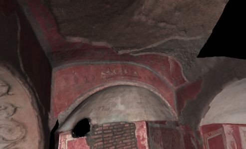
Il convegno LIMS 2018

Il convegno scientifico LIMS 2018, Luce, Imaging, Microscopia, Spettri di applicazione, http://www.frascati.
enea.it/LIMS2018/, si svolge presso il Centro Ricerche ENEA di Frascati il 17 e 18 maggio 2018 in occasione
dell’International Day of Light, https://www.lightday.org/, proclamato dall’UNESCO lo scorso novembre.
La data scelta è quella del 16 maggio, in ricordo della prima luce LASER ottenuta da Theodore H. Maiman il 16
maggio 1960. Lo scopo è di fornire ogni anno un appuntamento per illustrare con continuità il ruolo centrale che la
luce ha nella scienza, nella tecnologia, nella cultura e nell’educazione e, più in generale, nello sviluppo sostenibile
della società, con particolare attenzione rivolta ai giovani.
Il convegno LIMS e’ stato promosso per la prima volta dal Laboratorio Micro e Nanostrutture per la Fotonica
dell’ENEA C.R. Frascati ad ottobre 2015, in occasione dell’International Year of Light 2015 (www.frascati.enea.it/
LIMS2015/) nell’ambito di una serie di iniziative ENEA mirate alla diffusione e divulgazione dei temi della Luce
e delle tecnologie associate.
Sulla base del successo dell’edizione 2015, LIMS 2018 intende mettere in contatto ricercatori e tecnologi
provenienti da enti di ricerca, universita’, laboratori industriali e piccole e medie imprese della regione Lazio allo
scopo di presentare progetti e risultati significativi, scambiare conoscenze multidisciplinari e trasferire know-how
nel vasto campo delle tecnologie associate alla luce, favorendo l’integrazione di idee ed informazioni tra i diversi
ambiti di applicazione.
Il convegno ha ottenuto il patrocinio della SIF, Societa’ Italiana di Fisica, della SIOF, Societa’ Italiana di Ottica e
Fotonica, delle Università di Roma Sapienza, Tor Vergata, Roma Tre e Campus Biomedico, dell’INFN-LNF e del
CNR, i cui rappresentanti sono coinvolti nel Comitato Scientifico insieme all’ENEA.
E’ aperto sia alla vasta comunita’ scientifica degli addetti ai lavori, che ai numerosi operatori del settore ed alle
piccole e medie imprese laziali. Prevede uno spazio espositivo per poster ed una mostra tecnica riservata agli
operatori del settore.
La partecipazione, gratuita, offre un’occasione di incontro, di scambio e di approfondimento sui temi delle
tecnologie fotoniche e le loro prospettive di sviluppo, a partire dalle attività, i progetti e le competenze presenti
nelle sedi laziali dell’ENEA.
La Fotonica, una delle tecnologie abilitanti nel Programma Europeo H2020, fornisce oggi soluzioni in campi di
ricerca molto diversi fra loro e le Sessioni sono state articolate secondo le seguenti tematiche e applicazioni della
Fotonica:

 •   Sensoristica ottica, laser e spettroscopia per applicazioni biomedicali
 •   Tecnologie ottiche, laser e spettroscopia per applicazioni industriali
 •   Tecnologie ottiche, laser e materiali per l’energia
 •   Tecnologie laser, imaging e spettroscopia per l’ambiente, lo spazio ed i beni culturali
In questo quaderno-appunti sono raccolti i riassunti delle comunicazioni su invito e dei poster.
Insieme al Comitato Locale di LIMS 2018, con l’entusiasmo e l’interesse che la LUCE E LE TECNOLOGIE
BASATE SULLA LUCE meritano, auguro a tutti i partecipanti buon lavoro.

						                                                        Rosa Maria Montereali
SPONSOR e MOSTRA TECNICA

                                                                                              Crisel Instruments
                                                                                           Hamamatsu Photonics Italia
                                                                                                 Horiba Italia
                                                                                                  Optoprim

                                        Comitato Scientifico LIMS2018

                     Antonio Cricenti, ISM-CNR, Area della Ricerca di Roma Tor Vergata
                               Roberto Francini, Università di Roma Tor Vergata
                                        Augusto Marcelli, INFN - LNF
                                  Michele Marrocco, ENEA C.R. Casaccia
                            Francesco Michelotti, SAPIENZA Università di Roma
                             Rosa Maria Montereali (Chair), ENEA C.R. Frascati
                     Valentina Mussi, IMM-CNR, Area della Ricerca di Roma Tor Vergata
                          Emiliano Schena, Università Campus Bio-Medico di Roma
                                  Fabrizia Somma, Università di Roma Tre

                                           Comitato Organizzatore

                                 Francesca Bonfigli, ENEA C.R. Frascati
                                   Monica Cimino, ENEA C.R. Frascati
                                   Lori Gabellieri, ENEA C.R. Frascati
                                 Antonella Mancini, ENEA C.R. Frascati
                                   Flavio Miglietta, ENEA C.R. Frascati
                                Rosa Maria Montereali, ENEA C.R. Frascati
                                  Flaminia Rondino, ENEA C.R. Frascati
                                Maria Aurora Vincenti, ENEA C.R. Frascati

http://www.frascati.enea.it/LIMS2018/
Esposizione dell’artista Germana Brizio Relazioni su invito - LIMS2018
1. The discovery of gravitational waves and the contribution of optical technologies 10
Germana Brizio, artista versatile, ritrattista, scultrice ed incisore, nata nel 1953 a Ginosa Marina in provincia di 2. The making of molecular movies with femtosecond light flashes 12
Taranto, consegue la maturità al Liceo Artistico Statale di Lecce e completa gli studi all’Accademia di Belle Arti
di Roma con la tesi “La Luce nella Pittura di Caravaggio”. È abilitata all’insegnamento di Educazione Artistica e 3. Early Stage Diagnosis of Tumors and Diseases by Nanospectroscopy 14
Disegno e Storia dell’Arte, ha ampliato il suo percorso con esperienza di restauratrice e arte-terapeuta, formazione 4. Gold coated silicon nanowires for combined near-infrared photothermal treatment of cancer cells and
che ha arricchito lo spettro di conoscenza portandola ad esprimersi con una valenza artistica del tutto originale. La
sua pittura reinterpreta il rapporto “uomo-natura”, memore della lezione Caravaggesca, pittura che è incrocio di Raman monitoring of the process evolution 16
arte e scienza, dove un fascio di luce esce dalle tenebre della materia per farsi colore. 5. Transversal dose mapping and Bragg-curve reconstruction in proton-irradiated lithium fluoride detectors
Germana Brizio ha esposto dal 1973 in numerose sedi prestigiose, sia in Italia che all’estero e le sue opere by fluorescence microscopy 18
figurano in collezioni pubbliche e private, nazionali e internazionali. Tra le opere più pregiate figurano il ritratto
per Papa Francesco, il pannello pittorico (3x2) m2 “Stemma della Compagnia di Gesù” sulla facciata della Chiesa 6. Laser ablation for cancer removal: present and emerging solutions 20
di Sant’Ignazio di Loyola a Roma, l’opera pittorica su tela “Mater Poenitens” per la Chiesa del Sacro Cuore dei 7. Bloch surface wave biosensors for real-time study of fibronectin/phosphorylcholine-based biomedical
Gesuiti a Tirana, Albania. Molte sue opere e installazioni sono recensite su pregiate riviste e cataloghi specializzati coatings 22
nel settore, il suo nominativo è inserito nell’Enciclopedia di Arte Italiana Contemporanea (vol. IX) Casa Editrice
La Ginestra 1979, e nell’annuario di Art Diary Italia dal 1991. 8. Laser Induced Periodic Surface Structures: from physical phenomena to industrial applications 24
9. Quantum dots synthesis and laser patterning for light sources manufacturing 26
Durante il convegno scientifico LIMS 2018 (17 - 18 maggio 2018, ENEA C.R. Frascati), http://www.frascati.enea. 10. Innovative optical assessments for material investigation in automotive sector 28
it/LIMS2018/, vengono esposte due opere di Germana Brizio, pittura ad olio su tela, di espressione naturalistica
che indaga il rapporto luce colore. 11. Fluoride Crystals: High Performance Materials for Photonic Devices 30
12. Influence of Extreme Ultraviolet irradiation on structural properties of CVD grown graphene studied by
Raman mapping 32
13. Fiber Bragg Gratings for respiratory and relative humidity monitoring in biomedical applications 34
14. Optical properties and sensor applications of silver nanoparticles and nanoclusters 36
15. Exploiting quantum resources towards low-intrusive sample characterization 38
16. LINC: an interdepartmental laboratory at ENEA for femtosecond CARS spectroscopy 40
17. Transparent oxides for selective contacts and passivation in heterojunction silicon solar cells 42
18. 2D materials as promising strategy for enhanced efficiency and stability in perovskite photovoltaics 44
19. Matter structured by ultra-intense laser for conversion of concentrated solar energy 46
20. Review of ENEA activity in Concentrating Solar Power and new instruments developed for optical
diagnostics 48
21. Laser induced breakdown spectroscopy as diagnostic for tokamak and materials of fusionistic interest 50
22. Gamma radiation effects on materials and components investigated by optical characterization 52
23. EUV polarimetry in lab: multilayer characterization and phase retarder reflector development for space 54
24. Terahertz applications on art conservation at the ENEA Frascati 56
25. Laser-based scanners for high quality structural and colour digitalisation of medium/big Cultural
Heritage artworks 58
Poster
P1. Lithium fluoride luminescent detectors for X-FEL beam imaging 62
P2. Oxinitride coatings for aerospatial enviroments 63
P3. Research activity in the laboratory for inertial confinement fusion in ENEA - Centro Ricerche Frascati 64
P4. Enhancement of the laser ablation rate induced by oxides nanoparticles 65
P5. Laser photoacoustic spectroscopy for food fraud detection 66
P6. Atmospheric lidar for remote monitoring of natural hazards and early warning of forest fires 67
P7. Light filtering by thin-film coatings for an optical detection system 68
P8. Photoluminescence response of radiation-induced color centers in LiF crystals: the clinical dosimetry
challenge 69
P9. Remarkable performance of YBCO films through the inclusion of (Nb,Ta)-based efficient artificial
pinning centers for fusion application 70
P10. BaZrO3 inclusions in solution-derived YBa2Cu3O7-∂ epitaxial thin films studied by X-Ray Photoelectron
Spectroscopy 71
P11. Growth and characterisation of LiF films for low-energy proton beam diagnostics 72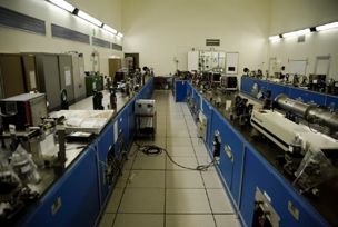
Workshop Luce, Imaging e Microscopia, Spettri di Applicazione - LIMS 2018
16:25 - 16:55 COFFEE BREAK - SESSIONE POSTER – MOSTRA TECNICA
ENEA C.R. Frascati, 17–18 maggio 2018
SESSIONE 4 : Sensoristica ottica, microscopia e spettroscopia per ambiente e biomedicale
Programma
GIOVEDI, 17 MAGGIO 2018 16:55 - 17:20 Daniela Lo Presti, Università Campus Bio-Medico di Roma
Fiber Bragg Gratings for respiratory and relative humidity monitoring in biomedical
08:30 REGISTRAZIONE applications
09:15 - 09:40 APERTURA LAVORI - Ing. A. Pizzuto, Dr.ssa R. Fantoni, Dr.ssa R.M. Montereali 17:20 - 17:45 Luca Burratti, Università di Roma Tor Vergata
Optical properties and sensor applications of silver nanoparticles and nanoclusters
SESSIONE 1 : Relazioni Generali 17:45 - 18:10 Emanuele Roccia, Università degli Studi Roma Tre
Exploiting quantum resources towards low-intrusive sample characterization
09:40 - 10:15 Pia Astone, SAPIENZA Università di Roma – INFN 18:10 - 18:35 M. Falconieri, ENEA C.R. Casaccia
The discovery of gravitational waves and the contribution of optical technologies LINC: an interdepartmental laboratory at ENEA for femtosecond CARS
spectroscopy
10:15 - 10:45 Tullio Scopigno, SAPIENZA Università di Roma
The making of molecular movies with femtosecond light flashes
VENERDI, 18 MAGGIO 2018
10:45 - 11:15 COFFEE BREAK - SESSIONE POSTER – MOSTRA TECNICA
08:30 REGISTRAZIONE
SESSIONE 2 : Laser, microscopia e spettroscopia per applicazioni medicali
SESSIONE 5 : Tecnologie ottiche, laser e materiali per l’energia
11:15 - 11:40 Antonio Cricenti, ISM-CNR
09:00 - 9:25 Francesca Menchini, ENEA C.R. Casaccia
Early Stage Diagnosis of Tumors and Diseases by Nanospectroscopy
Transparent oxides for selective contacts and passivation in heterojunction silicon solar cells
11:40 - 12:05 Annalisa Convertino, IMM-CNR
09:25 - 9:50 Antonio Agresti, Università di Roma Tor Vergata
Gold coated silicon nanowires for combined near-infrared photothermal treatment of cancer
cells and Raman monitoring of the process evolution 2D materials as promising strategy for enhanced efficiency and stability in perovskite
photovoltaics
12:05 - 12:30 Enrico Nichelatti, ENEA C.R. Casaccia
09:50 - 10:15 Daniele M. Trucchi, ISM-CNR
Transversal dose mapping and Bragg-curve reconstruction in proton-irradiated lithium
fluoride detectors by fluorescence microscopy Matter structured by ultra-intense laser for conversion of concentrated solar energy
12:30 - 12:55 Emiliano Schena, Università Campus Bio-Medico di Roma 10:15 - 10:40 Marco Montecchi, ENEA C.R. Casaccia
Laser ablation for cancer removal: present and emerging solutions Review of ENEA activity in Concentrating Solar Power and new instruments developed for
optical diagnostics
12:55 - 13:20 Alberto Sinibaldi, SAPIENZA Università di Roma
10:40 - 11: 05 Salvatore Almaviva, ENEA C.R. Frascati
Bloch surface wave biosensors for real-time study of fibronectin/phosphorylcholine-based
biomedical coatings Laser induced breakdown spectroscopy as diagnostic for tokamak and materials of
fusionistic interest
13:20 - 14:20 PRANZO
11:05 - 11:35 COFFEE BREAK - SESSIONE POSTER – MOSTRA TECNICA
SESSIONE 3 : Processi laser, materiali ed applicazioni industriali
SESSIONE 6 : Tecnologie ottiche ed imaging per lo spazio ed i beni culturali
14:20 - 14:45 Leonardo Orazi, Università di Modena e Reggio Emilia
11:35 - 12:00 Ilaria Di Sarcina, ENEA C.R. Casaccia
Laser Induced Periodic Surface Structures: from physical phenomena to industrial applications
Gamma radiation effects on materials and components investigated by optical characterization
14:45 - 15:10 Francesco Antolini, ENEA C.R. Frascati
12: 00 - 12:25 Paola Zuppella, IFN-CNR
Quantum dots synthesis and laser patterning for light sources manufacturing
EUV polarimetry in lab: multilayer characterization and phase retarder reflector development
15:10 - 15:35 Marie Marguerite Dugand, Centro Ricerche Fiat, Torino
for space
Innovative optical assessments for material investigation in automotive sector
12:25 - 12:50 E. Giovenale, ENEA C.R. Frascati
15:35 - 16:00 Mauro Tonelli, Università di Pisa
Terahertz applications on art conservation at the ENEA Frascati
Fluoride Crystals: High Performance Materials for Photonic Devices
12:50 - 13:15 Massimiliano Guarneri, ENEA C.R. Frascati
16:00 - 16:25 S. Botti, ENEA C.R. Frascati
Laser-based scanners for high quality structural and colour digitalisation of medium/big
Influence of Extreme Ultraviolet irradiation on structural properties of CVD grown graphene Cultural Heritage artworks
studied by Raman mapping
13:15 - 13.30 CHIUSURA LAVORI
Sessione inaugurale

                                   Relazioni Generali

   The discovery of gravitational waves and the contribution of optical technologies
                Pia Astone, SAPIENZA Università di Roma – INFN

             The making of molecular movies with femtosecond light flashes
                   Tullio Scopigno, SAPIENZA Università di Roma
Book of Abstracts

     1.   The discovery
     The discovery      of gravitational
                   of gravitational      waves
                                     waves  andand
                                                thethe contributionofofoptical
                                                    contribution        opticaltechnologies
                                                                                technologies

                                                                 *
                                                                                                                     2018
                                                     P. Astone

                                                                                                              Memo
                *
                    INFN, Sezione di Roma. Physics Department of the University “La Sapienza”
                                   Piazzale Aldo Moro 2, I-00185, Rome (Italy)

     Observations of compact binary coalescences by the Advanced LIGO and Virgo gravitational wave
     (GW) detectors have marked the beginning of a new era: first there were optical telescopes. Then
     radio wave detectors and X-ray observatories gave researchers a new method to view the cosmos.
     Now we can see it through GWs, and this allows us to explore new physics and astrophysics.
     The unprecedented sensitivity needed to unveil the faint GW signals has posed many technical
     challenges over the years: the detection concept is to measure the time it takes light to travel
     through the space distorted by GWs using precision interferometry. At each facility, laser light is
     injected into orthogonal tubes 3-4 km long. Mirrors at the ends of the tubes reflect the light back. A
     passing GW will increase the light travel time in one arm while decreasing it in the other. This time
     difference is converted into a change in intensity and measured on photodetectors.
     The discovery and the associated potentialities are such that the Nobel Prize in Physics was, in the
     year 2017, awarded to three fathers of these experiments. I will present the results achieved so far,
     describing the fundamental role of optical technologies, together with the plans to improve the GW
     network sensitivity in the upcoming years.

                        Figure 1 The Virgo gravitational wave interferometer (at EGO, Cascina)

     *Corresponding author: pia.astone@roma1.infn.it

10                                                                                                                                              11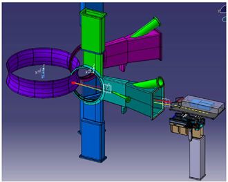
Book of Abstracts

     2.   The making of molecular movies with femtosecond light flashes

                                                                                                                      2018
             The making of molecular movies with femtosecond light flashes

                                                T. Scopigno1
                         1
                             Dipartimento di Fisica, Università Roma “Sapienza”                                Memo
 Visualizing the evolution of molecular structures through their transition states is the key to
 understand elemental physical processes, chemical reactions and, ultimately, biological function.
 The task is hampered by the simultaneous need of structural and temporal resolutions adequate at
 the atomic scale. The challenge can be embraced with different approaches. Here I will discuss how
 to reach the goal working on the potential of an old technique, the Raman effect. Using ad-hoc
 designed sequences of ultrashort optical pulses allows first stimulating and subsequently detecting
 inter-atomic vibrations via the coherent version of the Raman effect, enabling unprecedented
 temporal precision combined with structural resolution [1]. This ultimately allows recording frames
 of atomic dynamics which can be used for the making of molecular movies [2].

 [1] G. Batignani, D. Bossini, N. Di Palo, C. Ferrante, E. Pontecorvo, G. Cerullo, A. Kimel and T. Scopigno.
 Nature Photonics, 9, 506 (2015).
 [2] C. Ferrante, E. Pontecorvo, G. Cerullo, M. H. Vos, T. Scopigno, Nature Chemistry 8, 1137 (2016).

 *Corresponding author: tullio.scopigno@roma1.infn.it

12                                                                                                                                               13
Book of Abstracts
3. Early Stage
Early Stage Diagnosis
Diagnosisofof
Tumors andand
Tumors Diseases by Nanospectroscopy
Diseases by Nanospectroscopy
A. Cricenti 2018
Istituto di Struttura della Materia (ISM-CNR)
Via del Fosso del Cavaliere 100, Rome, 00133, Italy
Memo
Keywords: (Raman, SNOM, IR, Cancer Cells, ALS)
Carcinomas are complex biochemical systems and in the past their diagnosis was based on
morphological differences between malignant cells and their benign counterpart. Recently the
paradigm has changed and great interest is focused now on the biochemical profile of tumours
in view of the availability of new drugs that specifically target neoplastic cells. This new
paradigm requires biochemical analysis of each tumour in order to establish the correct
personalized oncological “target therapy”. Understanding the mechanism of molecular
alterations of a specific tumour is a critical issue to prognosticate its behaviour and to predict
the response to personalized therapy. Raman spectroscopy (RS) is a non-invasive optical
label-free tool increasingly used to get molecular fingerprints of biological tissues. It is able to
provide bioanalytical information on any molecule with high specificity. Technological
advances over the last decade have created a new and faster Raman imaging microscope
instrument, providing morphological tissue investigation of large areas, coupled with point-
by-point spectral analysis of biochemical composition. This option is important not only for
discrimination between healthy and pathological tissues, but especially for pre-cancerous
tissue state earlier detection and understanding. Raman mapping of biological tissues have
shown that the microscope can operate at a few micron resolution, in order to distinguish
between healthy and malignant tissues [1]. The potential of IR spectroscopy to characterise
cancerous tissues has long been recognised and studies of various cancers by many groups
have established that regions of malignant tissue can be easily identified on the basis of its IR
spectrum. Early diagnosis of cancer requires an instrument providing specific chemical
images at sub-cellular level and the development of diagnostic imaging. A SNOM meets
these requirements provided that it can been coupled with an appropriate infrared light source,
that can be based on Free Electron Laser, femtosecond laser or quantum cascade laser [2].
We present Raman and Infrared Scanning Near-field Optical Microscopy (SNOM) in their
spectroscopic mode, that is related to the local chemical composition and, thus, to the
biological properties of the sample, for tissue imaging and early cancer diagnostics.
Applications in the case of Oesophagous [3] and Cervical Cancer [4] as well as in the
progression of Amyotrophic Lateral Sclerosis (ALS) will be presented.
[1] Çulha M, Bioanalysis, 7, 2813 (2015)
[2] Cricenti A, Luce M, Tolk NH, Margaritondo G; Nanosci. Nanotechnol. Lett.; 3 (2011) 913;
[3] Smith AD, Siggel-King MRF, Holder GM, Cricenti A, Luce M, Harrison P, Martin DS, Surman
M, Craig T, Barrett SD, Wolski A, Dunning DJ, Thompson NR, Saveliev Y, Pritchard DM, Varro A,
Chattopadhyay S, Weightman P; Applied Physics Letters; 102 (2013) 053701.
[4] Halliwell Diane E, Morais Camilo LM, Lima Kássio MG, Trevisan Julio, Siggel-King Michele RF,
Craig Tim, Ingham James, Martin David S, Heys Kelly A, Kyrgiou Maria, Mitra Anita, Paraskevaidis
Evangelos, Theophilou Georgios, Martin-Hirsch Pierre L, Cricenti Antonio, Luce Marco, Weightman
Peter, Martin Francis L; Scientific Reports; 6 (2016) 29494.
*Corresponding author: antonio.cricenti@ism.cnr.it
14 15Book of Abstracts
4.Array of disordered
Gold silicon
coated silicon nanowires
nanowires for coated by anear-infrared
combined gold film for photothermal
combined NIR
treatment of cancer cells and Raman monitoring of the process evolution
photothermal treatment of colon cancer cells and Raman monitoring of the
process evolution
2018
A. Convertino1*, M. Mussi1, L Maiolo1, M. Ledda2, M. G. Lolli2, G. Fortunato3, Memo
Massimiliano Rocchia4, Antonella Lisi2
1
Institute for Microelectronics and Microsystems, CNR, 00133 Rome, Italy
2
Institute of Translational Pharmacology, CNR, 00133 Rome, Italy
3
Institute for Microelectronics and Microsystems, CNR, 95121 Catania, Italy
4
Thermo Fisher Scientific, 20090 Rodano, Italy
Photothermal therapy (PTT) assisted by nanomaterials is a promising minimally invasive alternative
to the traditional cancer surgery. Here, we explore the PTT properties of a gold based
nanostructured platform suitable to be directly integrated in fiber laser systems rather than injected
into the human body as occurring for the most reported PTT nanoagents. In particular, the
phothermal properties of an array of disordered silicon nanowires coated by a thin gold film
(Au/SiNWs) were tested on a monolayer of human colon adenocarcinoma cells (Caco-2) irradiated
with a 780 nm laser. Au/SiNWs allowed an efficient photothermal action and simultaneous
monitoring of the process evolution through the Raman signal coming from the irradiated cellular
zone. Strong near infrared (NIR) absorption, overlapping three biological-windows, cell-friendly
properties and effective fabrication technology make Au/SiNWs suitable both to be integrated in
surgical laser tools and as in vitro platform to develop novel PTT protocols using different cancer
types and NIR sources.
Figure 1 Working principle of Au/SiNWs as photothermal platform
*Corresponding author: annalisa.convertino@cnr.it
16 17Book of Abstracts
5. Transversal
Transversal dose dose mapping
mapping and Bragg-curve
and Bragg-curve reconstruction
reconstruction in proton-
in proton-irradiated
irradiated lithium fluoride detectors by fluorescence microscopy
lithium fluoride detectors by fluorescence microscopy 2018
E. Nichelatti1*, M. Piccinini2, A. Ampollini2, L. Picardi2, C. Ronsivalle2, F. Bonfigli2,
M.A. Vincenti2, R.M. Montereali2
Memo
1
ENEA C.R. Casaccia, Fusion and Technologies for Nuclear Safety and Security
Via Anguillarese 301, S. Maria di Galeria, Rome, 00123, Italy
2
ENEA C.R. Frascati, Fusion and Technologies for Nuclear Safety and Security
Via E. Fermi 45, Frascati (RM), 00044, Italy
When lithium fluoride (LiF) is irradiated with a suitable kind of ionising radiation (�- or X-rays,
ions, electrons, extreme UV light, etc.), lattice defects, known as colour centres (CCs), form in the
crystalline structure. Some of them luminesce in the visible when illuminated by blue light, even at
room temperature. This peculiarity is here exploited for imaging in a fluorescence microscope the
photoluminescence (PL) transversal maps and longitudinal distributions due to CCs formed in LiF
detectors placed along the path of proton beams, with energies from 3 to 35 MeV, delivered by a
linear accelerator for protontherapy applications that is under construction at ENEA C.R. Frascati
(TOP-IMPLART project). The recorded PL images are elaborated to reconstruct the 2D cross
sections [1] and depth distributions (Bragg curves) [2] of the absorbed dose. In this way, useful
pieces of information are obtained regarding the kinematics of CC formation in LiF and for
advanced proton-beam diagnostics.
Figure 1 Irradiation of a LiF crystal with 18 MeV protons. Calculated dose map (left) as obtained from the
recorded transversal PL image; comparison between measured and simulated PL depth distributions (right).
The latter ones correspond to the proton Bragg curve in the material.
[1] M. Piccinini et al., Europhys. Lett. 117, 37004 (2017)
[2] E. Nichelatti et al., Europhys. Lett. 120, 56003 (2017)
*Corresponding author: enrico.nichelatti@enea.it
18 19Book of Abstracts
6. Laser
Laser ablation
ablation forfor cancer
cancer removal:
removal: present
present and emerging
and emerging solutions
solutions
E. Schena1*, M. A. Caponero2 2018
Memo
1
Università Campus Bio-Medico di Roma, Rome, Italy
2
Research Centre of Frascati, ENEA, Rome, Italy
Laser ablation (LA) has gained broad clinical acceptance, and it is particularly attractive as an
alternative to surgical resection for patients who are not good surgical candidates. LA is performed
by guiding the laser light via a thin and flexible optical fiber, hence the treatment is minimally
invasive techniques and can be performed under Endoscopic ultrasound-guidance via either a
percutaneous approach or via anatomical ducts. It offers several benefits for patients, as the
reduction of operative trauma, adhesions and wound dehiscence, reduction of recovery time and of
the incidence of post-surgical complications.The selectivity of the treatment is a key asset to obtain
an optimal clinical outcome. Three promising solutions for improving the selectivity are the real-
time temperature monitoring, the use of models to plan a patient-specific treatment, and the use of
highly absorbing nanoparticles to improve the selectivity of the procedure.This seminar will discuss
the factors which influence LA outcome, and if and how the mentioned emerging solutions may
significantly improve the clinical outcomes of laser ablation. Attention will be devoted to the use of
fiber Bragg grating (FBGs) sensors for temperature monitoring during LA. This solution allows
performing highly resolved temperature monitoring by inserting a single, small-sized needle
embedding several FBGs inside the organ [1].
Figure 1 Reconstructed Computed Tomography image showing the optical applicator and the two probes
embedding FBG sensors. A three-dimensional temperature map reconstructed by FBGs measure is shown.
[1] P Saccomandi, E Schena, MA Caponero et al. Theoretical analysis and experimental evaluation of laser-
induced interstitial thermotherapy in ex vivo porcine pancreas. IEEE Trans Bio-Med Eng 2012; 59:2958-
2964.
*Corresponding author: e.schena@unicampus.it
20 21Book of Abstracts
7. Bloch surface
Bloch surface wave biosensors
wave biosensors for real-time
for real-time study
study of offibronectin/
phosphorylcholine-based biomedical coatings
fibronectin/phosphorylcholine-based biomedical coatings 2018
A. Sinibaldi1*, V. Montaño-Machado2, N. Danz3, P. Munzert3, F. Chiavaioli4, D. Mantovani2, and
Memo
F. Michelotti1
1
Department of Basic and Applied Science for Engineering, SAPIENZA University of Rome, Italy.
2
Laboratory for Biomaterials and Bioengineering (CRC-I), Dept. of Min-Met-Materials Eng. &
CHU de Quebec Research Center, Laval University, Quebec City, Canada.
3
Fraunhofer Institute for Applied Optics and Precision Engineering IOF, Jena, Germany.
4
Nello Carrara Institute of Applied Physics IFAC, Florence, Italy.
The characterization of the interaction of proteins with surfaces is of high relevance to understand
the biological performance of biomaterials. Special complexity is presented when characterizing
coatings of molecules with high difference in molecular weight as fibronectin (FN, 440 kDa) and
phosphorylcholine (PRC, 181 Da), biomolecules largely studied for cardiovascular applications to
improve endothelialization and hemocompatibility, respectively [1,2]. In the present work, a
combined label-free and fluorescence optical technique was used to study FN-PRC coatings created
through the combination of adsorption and grafting processes. To accomplish this, one dimensional
photonic crystals (1DPCs) supporting Bloch surface waves (BSW) [3] were interrogated in label-
free and enhanced fluorescence operation modes. The label-free mode was obtained exciting a BSW
by means of a prism coupler in the Kretschmann-Raether configuration producing a dip in the
reflectance; by tracking such a minimum position, it is possible to monitor changes in the refractive
index as well as molecular interactions at the surface. Moreover, the enhanced fluorescence mode
offers the possibility to confirm the presence of protein levels evaluated in label-free experiments
with a sharp improvement of the resolution of the technique [4]. In conclusion, such a combined
technique could provide data - in real time - that are relevant for the further understanding of the
biological performance of coatings for medical devices.
[1] A. L. Lewis, et al., Biomaterials 21, 1847–1859 (2000).
[2] G. Li, et al., Colloids Surfaces B: Biointerfaces 81, 255–262 (2010).
[3] A. Sinibaldi, et al., Analytical and Bioanalytical Chemistry 407, 3965–3974 (2015).
[4] A. Sinibaldi, et al., Biosensors and Bioelectronics 92, 125–130 (2017).
*Corresponding author: alberto.sinibaldi@uniroma1.it
22 23Book of Abstracts
8. LaserInduced
Laser InducedPeriodic
PeriodicSurface
SurfaceStructures:
Structures:from
fromphysical
physicalphenomena
phenomena to
to
industrial applications
industrial applications 2018
1
L. Orazi 1*
Memo
University of Modena and Reggio Emilia
Laser Induced Periodic Surface Structures (LIPSS) were been for years a topic of great interests for
physicist. Although a 50 years long history [1] the models to explain the underlying phenomena are
not completely clear and are debated by scientific community. On the same time the diffusion of
new, reliable and less expensive ultrashort pulsed laser permits to substantially increase the treated
area and the regularity of obtained micro- and nano-structures [2]. These factors result in new
possible applications of LIPSS based surface structuring like friction reduction for tribology [3],
wettability control [4][5], plasmonic [6] and biomedical [7]. This contribution will face the new
advances on the basis of the last results presented in literature.
Figure 1 Left) LIPSS on stainless steel for tribology applications. Mid) hydrophobic structures on nickel
plate. Right) Light diffraction for aesthetic/anti-counterfeiting applications
[1] Milton Birnbaum, “Semiconductor Surface Damage Produced by Ruby Lasers,” J. Appl. Phys., vol. 36,
no. 11, (1965).
[2] I. Gnilitskyi, T. J.-Y. Derrien, Y. Levy, N. M. Bulgakova, T. Mocek, and L. Orazi, Sci. Rep., vol. 7, no.
1, (2017).
[3] I. Gnilitskyi, I. Pavlov, F. Rotundo, L. Orazi, C. Martini, and F. O. Ilday, in Conference on Lasers and
Electro-Optics Europe - Technical Digest, vol. 2015-August (2015).
[4] L. Orazi, I. Gnilitskyi, and A. P. Serro, J. Micro Nano-Manuf., vol. 5, no. 2, (2017).
[5] L. Orazi, I. Gnilitskyi, I. Pavlov, A. P. Serro, S. Ilday, and F. O. Ilday, CIRP Ann. - Manuf. Technol.,
vol. 64, no. 1 (2015).
[6] I. Gnilitskyi, S. Mamykin, M. Dusheyko, T. Borodinova, N. Maksimchuk, and L. Orazi, Frontiers in
Optics, p. JW4A.88 (2016).
[7] I. Gnilitskyi, M. Pogorielov, D. Dobrota, R. Viter, L. Orazi, and O. Mischenko, Conference on Lasers
and Electro-Optics (2016), p. AW4O.6 (2016).
*Corresponding author: leonardo.orazi@unimore.it
24 25Book of Abstracts
Quantum
9. Dots dots
Quantum Synthesis andand
synthesis Laser
laserPatterning
patterning for
for Light Sources
light sources Manufacturing
manufacturing
F. Antolini1*, I.D.W.Samuel2, G. Raciukaitis3
2018
1
ENEA, Fusion and Nuclear Security Dept., Photonics Micro and Nanostructures Laboratory,
Memo
Via E. Fermi 45, 00044 Frascati (Rome), Italy
2
Organic Semiconductor Centre, SUPA, School of Physics and Astronomy,
University of St Andrews, St Andrews, KY16 9SS, U
3
Center for Physical Science and Technology Department of Laser Technologies,
Savanoriu Av. 231, LT02300, Vilnius Lithuania
The synthesis of colloidal photo-luminescent semiconductor, i.e. CdS and CdSe QDs (figure 1),
from single source precursors [1] is presented both in solution and in the form of thin films. These
nanomaterials received a considerable interest within the material science, because together with
polymers are one of the key steps for new photonic devices manufacturing.
The implementation of direct laser patterning (DLP) of nanoparticles is also illustrated as a suitable
alternative for the fabrication of hybrid organic/nanoparticles based optical devices [2]. DLP
techniques do not require complex laser systems or the use of dangerous chemical post treatments
so they can be of advantage in such as Organic Light Emitting Diodes (OLEDs) manufacturing.
Figure 1 Semiconductor QDs showing different emission colors (from red to green)
obtained just by changing the reaction time of the synthesis.
[1] M.Z. Malik, M. Afzaal, P. O’Brien, Chem. Rev. 110, 4417 (2010)
[2] A. K. Bansal, M. T. Sajjad, F. Antolini, L. Stroea, P. Gečys, G. Raciukaitis, P. André, A. Hirzer,
V. Schmidt , L. Ortolani, S. Toffanin, S. Allard, U. Scherf & I. D.W. Samuel, Nanoscale 7, 11163, (2015)
*Corresponding author: francesco.antolini@enea.it
26 27Book of Abstracts
10. Innovative
Innovative optical
optical assessments
assessments forfor materialinvestigation
material investigation in
inautomotive
automotivesector
sector
2018
Marie Marguerite Dugand, Nello Li Pira, Luca Belforte
Centro Ricerche FIAT – Gml Physical Analysis – Torino (Italia) Memo
CRF – Group Materials Labs are involved in the qualification and definition of the specifications
concerning automotive materials and specifically in the Physical Analysis department the
mission is:
• Innovations in Optical & Electronics materials and assessment
• Development of innovative functional surfaces (e.g. antireflective, hydro-oleo-phobic..)
• Display qualification (optical, mechanical)
• Photo-Colorimetric assessment
• Integration of electronics for novel touch surfaces
• Morphology analysis and interaction with optical output
Functional surfaces need protective cover lenses with antiglare, anti-scratch, anti-abrasion,
durability against UV and chemicals attacks characteristics. CRF initially defined the antiglare
threshold effect on the external coating that covers the display: the reason is to avoid any internal
reflection which could affect the display visibility. Concerning the anti-scratch and anti-
abrasion characteristics, durability against UV and chemicals attacks, GML labs performed all
the test to evaluate the treatments on the cover lenses and the Optical Lab performed all
measurement before and after the treatments with Spectrophotometers, Spectroradiometers and
Gonio reflectometer to check and measure the Optical differences. Other topics to investigate are
the milky effect on whole surface due to external roughness of the antiglare coating and the
Birefringence, the optical delay generated by internal stresses due to the manufacturing
processes.
Examples and Theory will be presented.
*Corresponding author : mariemarguerite.dugand@crf.it , nellolipira@crf.it , lucabelforte@crf.it
28 29Book of Abstracts
11. Fluoride
FluorideCrystals:
Crystals:High
HighPerformance Materials
Performance for Photonic
Materials Devices
for Photonic Devices
Mauro Tonelli 1,2*
2018
Memo
1
Dipartimento di Fisica, Universita’ di Pisa, Italy
2
Nest-Nanoscience Insitute, CNR, Pisa, Italy
We developed high-quality single fluoride crystals grown by Czochralski technique. This activity
covers different applications such as IR and visible laser, RX detection, metrologic applications and
solid state crycooler.
The active parts of these devices are insulator hosts containing fluorine doped with trivalent rare
earths to develop solid state laser in the UV, visible and near infrared wavelength region. We have
investigated the spectroscopic properties and energy transfer mechanisms to tune high-efficiency IR
tunable lasers (about 300 nm) in CW and pulsed operation regime. By the same materials we have
investigated laser emission in the visible region by fluoride crystal doped with Pr3+ and Dy3+ and
showed the potential applications for optical atomic clock. It has been showed for the first time the
yellow CW laser emission by fluoride crystals.
Moreover, we have investigated the cooling effect on insulator materials by optical pumping. In
particular we evaluated the critical cooling parameters and EQE (ext) of different crystals. We
studied LiYF4 crystals doped with different Yb3+ doping levels. Moreover, it has been estimated
the cooling power for possible optical crycooler applications. On the same materials (LiYF4) we
proposed a new possible scheme to increase the cooling efficiency. This has been possible co-
doping the samples by Yb3+ and Tm3+. This approach has allowed to obtain the lowest temperature
(87 K) and the highest T=190 K.
*Corresponding author: mauro.tonelli@unipi.it
30 31Book of Abstracts
12. Influence
Influence of Extreme
of Extreme Ultraviolet
Ultraviolet irradiation
irradiation onon structuralproperties
structural properties of
of CVD
CVD
grown graphene studied by Raman mapping
grown graphene studied by Raman mapping 2018
S. Botti1*, L. Mezi1, A. Rufoloni1, A. Vannozzi 1,
Memo
S. Bollanti1, F. Flora1, S. Gay2
1.
Department of Fusion and Technologies for Nuclear Safety and Security, ENEA Via E. Fermi, 45,
00044 Frascati, Italy
2.
Horiba Italia srl, Via L. Gaurico, 209, 00143 Rome, Italy
The Extreme Ultra-Violet (EUV) irradiation influence on structural properties of graphene was
studied by Raman mapping, AFM and SEM. CVD grown mono-layer graphene films
(Graphenea) were irradiated at four different doses by using the ENEA (FSN-FUSPHY-SAD)
Discharge Produced Plasma (DPP) EUV source. The DPP emits more than 30 mJ/sr/ pulse on the
10-18 nm wavelength band (69-124 eV).
The experimental data suggest that under EUV irradiation defects were generated in the carbon
matrix due to the breaking of sp2 bonds with a subsequent oxidation in the DPP vacuum chamber
residual atmosphere. The oxidation leads to the formation of cracks and holes, similarly to that
happens with thermal oxidation but only on the irradiated areas.
The EUV-induced oxidation of graphene provides a possible route to graphene patterning.
*Corresponding author: sabina.botti@enea.it
32 33Book of Abstracts
13. Fiber
FiberBragg
BraggGratings
Gratingsfor
forrespiratory
respiratoryand
andrelative
relativehumidity
humidity monitoring
monitoring in
biomedical applications
biomedical applications 2018
1
D. Lo Presti1*, M.A. Caponero2, R. D’Amato2
Memo
Università Campus Bio-Medico di Roma - Rome, Italy
2
ENEA Research Centre of Frascati – Frascati (Rome), Italy
Photonics is driving innovations across several fields, spreading its wings from telecommunications
to human healthcare monitoring. Among fiber optic sensors, fiber Bragg gratings (FBGs) are the
most widespread in biomedical applications. They are characterized by low signal loss, EMI
immunity, small size, light weight and good metrological properties combined with the
multiplexing ability which allows spatial resolution of a variety of measurements. The popularity of
FBGs in medicine is mainly due to the development of wearables to monitor vital signs (e.g., tidal
volume, respiratory frequency, and heart rate) also in harsh environments (e.g., during Magnetic
Resonance exam). In addition, FBGs are used for chemical sensing with particular regard to relative
humidity (RH). FBGs sensitivity to RH is often achieved by coating the grating with hygroscopic
materials which react to moisture’s air content changes. This presentation aims to underline the
FBGs benefits for working in biomedical field focusing on two specific applications: i) the
respiratory monitoring by wearables to detect abnormal changes of respiratory parameters which
can be a predictor of physiological dysfunctions [1], and ii) the RH monitoring during mechanical
ventilation to improve the humidification process of delivered gases [2].
Figure 1 FBG-based systems for respiratory and RH monitoring.
[1] C. Massaroni, D. Lo Presti et al. (2018). Smart textile for respiratory monitoring and thoraco-abdominal
motion pattern evaluation, Journal of Biophotonics;
[2] C. Massaroni, D. Lo Presti et al. (2017). Fiber Bragg grating probe for relative humidity and respiratory
frequency estimation: assessment during mechanical ventilation. IEEE Sensors Journal.
* Corresponding author: d.lopresti@unicampus.it
34 35Book of Abstracts
14. Optical properties
Optical properties and sensor
sensor applications
applications ofofsilver
silvernanoparticles
nanoparticlesand
and
nanoclusters
nanoclusters 2018
L. Burratti1,*, F. De Matteis1,2, F. Mochi1,2, R.Francini1,2, M. Casalboni1,2, E. Bolli1, P. Prosposito1,2
1
Memo
Industrial Engineering Department and INSTM, University of Rome “Tor Vergata”, Via del
Politecnico 1, 00133, Roma, Italy
2
Center for Regenerative Medicine, University of Rome “Tor Vergata”, Via Montpellier 1, Rome,
00133, Italy.
Optical properties of metal nanoparticles depend on the type of metal and on the size [1]. When an
electromagnetic wave interacts with a colloidal suspension of metal nanoparticles (MNPs), an
optical response appears depending on MNPs dimensions. If the average diameter is in the range 2-
10 nm we can talk of nanoparticles and the physical phenomenon occurring in presence of light is
the Surface Plasmon Resonance (SPR). In this case, electrons can oscillate on the surface of
particles in resonance with the characteristic incident wavelength. When the average size of the
colloids falls below 2 nm, peculiar properties of nanoparticles disappear and molecules-like
electronic levels appear generating absorption and radiative decay process (luminescence). These
structures are called metal nanoclusters (MNCs). We synthesized both silver nanoparticles and
silver nanoclusters and we tested these systems as heavy metal ions optical detector [2] measuring
absorption and emission spectra in absence and in presence of ionic pollutants. The MNPs system
showed a good sensibility and selectivity to the presence of Ni2+ and Co2+. We synthesized also two
different luminescent silver NCs exploiting two capping agents: L-glutathione (GSH) and
poly(methacrylic acid) (PMAA). We tested also these two systems as optical detectors following
the changes induced in the absorption and luminescence bands.
a) b)
Figure 1. Photographs of different samples: (1) AgNPs, (2) AgNCs with GSH as capping agent, (3) AgNCs
with PMAA; under visible light (a) and UV irradiation (b). Both solutions of AgNCs show luminescence
emission, while AgNPs do not present a quantum behavior.
[1] I.Diez, R. H. A. Ras, Flourescent silver nanoclusters, Nanoscale 3.5, 1963-1970 (2011).
[2] P. Prosposito, F. Mochi, E. Ciotta, M. Casalboni, F. De Matteis, I. Venditti, L. Fontana, G. Testa, I.
Fretoddi, Hydrophilic silver nanoparticles with tunable optical properties: application for the detection of
heavy metals in water, Beilstein journal of nanotechnology 7 1657-1661 (2016).
*Corresponding author: Luca.Burratti@uniroma2.it
36 37Book of Abstracts
15. Exploiting quantum resources towards low-intrusive sample characterization
Exploiting quantum resources towards low-intrusive sample characterisation
E. Roccia1*
2018
1
Università degli studi Roma Tre, Dipartimento di scienze, Via della Vasca 84, 00146 Roma.
Memo
The identification of genuine properties of single molecules as well as their detection in relevant
environments, as in the case of aqueous solutions or in vivo conditions for biological interests,
represents an ultimate goal for physical and chemical analysis. On the way through, the
development of new approaches for non-destructive detection becomes a necessary and appealing
requirement. In this context quantum resources-based schemes show themselves as attractive tools
as their use allows the overcome of limitations characterising conventional counterparts, such as
high-power damages, fundamental issue for biological purpose. Purely quantum coherence is the
main character to be considered in performing low-intrusive quantum measurements. Quantum
metrology [1] is presented as the proper framework to fully harnessing its potentiality, opening the
perspective towards spectroscopies that make use of quantum probes. Due to their paramount
importance, chiral solutions should be good candidate for such an investigation. A standard way to
characterise the chirality of a specimen relies on light-matter interaction-based techniques [2,3],
being the optical activity a manifestation of this property: due to the optical rotatory power, the
polarisation plane changes once linearly polarised light passes throughout the sample. Optical
rotatory dispersion is finding renewed interest in the light of quantum resources-based protocols of
metrology [4]. For these purposes, exploiting quantum correlations, e.g. in the form of
entanglement, classical boundaries can be exceeded, thus ensuring a more efficient estimation of
phase and de-phasing in polarisation-based experiments. Implementation of a multi-parameter
estimation of phase and de-phasing, instead measuring these quantities separately, can improve the
result [5-6].
[1] V. Giovannetti et al., Science 306, 1330-1336 (2004).
[2] L.D. Barron et al., Chemical Physics Letters 492, 199-213 (2010).
[3] Y. He et al., Applied spectroscopy 65, 699-723 (2011).
[4] N. Tischler et al., Science Advances 2, e1601306 (2016).
[5] M.D. Vidrighin et al., Nature Communications 5, 3532 (2014).
[6] E. Roccia et al, Quantum Science and Technology 3, 01LT01 (2018).
*Corresponding author: emanuele.roccia@uniroma3.it
38 39Book of Abstracts
16.LINC:
LINC:an an interdepartmentalLaboratory
Interdepartmental laboratory at
atENEA
ENEAfor
forFemtosecond
femtosecond CARS
CARS
spectroscopy
Spectroscopy 2018
M. Falconieri1, S. Gagliardi1, M. Marrocco2, C. Merla3, F. Rondino4
1
ENEA FSN-TECFIS, 2ENEA DTE-PCU, 3ENEA SSPT-TECS
Memo
C.R. Casaccia via Anguillarese 301, 00123 Rome, Italy
4
ENEA FSN-TECFIS, C.R. Frascati, Via Enrico Fermi, 45, 00044 Frascati (RM), Italy
Imaging techniques based on vibrational spectroscopy are widely recognized tools for identification
and localization of compounds in a broad range of fields, from biology to materials science and
chemistry. In particular, third-order coherent techniques, such as coherent anti-Stokes Raman
scattering (CARS), have gained a wide interest [1] because of improved sensitivity and imaging
performances in comparison to their spontaneous counterpart. Exploitation of such capabilities in
the frame of ENEA activities is desirable and requires merging different skills and dedicated
instrumentation. Here, we present an initiative for the joint interdepartmental realization in the
ENEA Casaccia research center of a micro-CARS apparatus based on a femtosecond laser
oscillator. The Project leverages on the presence in the FSN-TECFIS laboratories of a fully
implemented femtosecond laser, thus allowing the realization of the micro-CARS system with
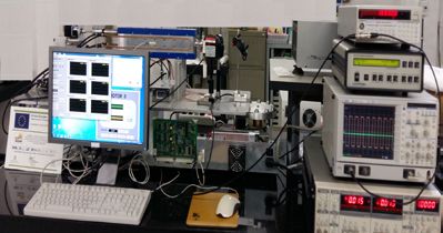
limited investments. The key feature of the setup, shown in fig.1, is the use of the supercontinuum
generated in a photonic crystal fiber as Stokes beam [2]; a home-built multiphoton microscope (not
shown in the figure) enables sample mapping by detecting both forward- and epi- signals. Presently,
the experimental setup is running and its performances are being assessed on test samples [3].
Figure 1 Layout of the micro-CARS experimental setup. Only the forward-CARS detection is shown.
[1] J-X. Cheng and X. S. Xie, J. Phys. Chem. B 108, 827 (2004)
[2] T. W. Kee and M. T. Cicerone, Opt. Lett. 29 (23), 2701 (2004)
[3] M. Falconieri, M. Marrocco, C. Merla, S. Gagliardi, F. Rondino, ECONOS 2018, 17th European
Conference On Non-Linear Optical Spectroscopy (2018)
*Corresponding author: mauro.falconieri@enea.it
40 41Book of Abstracts
17. Transparent
Transparentoxides
oxidesfor
forselective
selectivecontacts
contactsand
andpassivation
passivation in
in heterojunction
heterojunction
silicon solar cells
1*
silicon solar cells
1,2
F. Menchini , L. Serenelli , L. Martini , M. Izzi and M. Tucci 1,2 1 1
2018
Memo
1
ENEA C.R. Casaccia, Photovoltaic Technologies Laboratory (DTE-FSN-TEF), Rome
2
Sapienza University, Department of Information Engineering, Electronics and
Telecommunications (DIET), Rome.
Sunlight is by far the most abundant natural source of energy, which could cover the needs of all
mankind. Photovoltaic cells are the only technology able to directly convert the solar radiation into
available electric power. The management of light within solar cells has always been a central topic
in the photovoltaics optimization process. In particular, the transparency of the top layers of the
cells strongly influence the absorption of light in the active medium and thus the device
performances. Indeed in highly-efficient silicon heterojunction solar cells, the top of the cell is
usually constituted by a thin layer of amorphous silicon (a-Si:H) for passivation and charge
collection, and a transparent conductive oxide layer with a metal grid for extraction. Despite its very
good surface passivation capabilities, a-Si:H is not thermally stable and introduces undesired
parasitic absorption in the ultra-violet (UV) region of the solar spectrum.
In this presentation we show the results of studies aimed at increasing the absorption inside silicon
by substituting the a-Si:H layer with a combination of an amorphous silicon oxide layer (a-SiOx:H)
for buffer passivation and a high work function transparent oxide layer, such as Molybdenum
Oxyde (MoOx), as hole extractor. We show a net increase in the generated current in the 350-600
nm wavelength range due to increased transparency of the front layers.
1.0
0.8
N orm a liz e d IQ E
0.6
0.4
0.2 C ells ba s ed on
a -S i:H
a -S iO x :H /MoO x
0.0
400 500 600 700 800 900
W a ve leng th (nm )
Figure 1 Normalised Internal Quantum Efficiency of heterojunction solar cells
based on a-Si:H (red) and on a-SiOx:H/MoOx (blue).
*Corresponding author: francesca.menchini@enea.it
42 43Book of Abstracts
18. 2D
2Dmaterials
materials as
as promising
promising strategy
strategy for
for enhanced
enhanced efficiency
efficiencyand
andstability
stabilityin
perovskite photovoltaics
in perovskite photovoltaics 2018
A. Agresti1*, S. Pescetelli1, F. Bonaccorso2, Y. Busby3, A. Vinattieri4, D. Catone5 and A. Di Carlo1
1
C.H.O.S.E. (Centre for Hybrid and Organic Solar Energy), Electrical Engineering Department,
Memo
University of Rome Tor Vergata, Via del Politecnico 1, I-00133 Rome, Italy.
2
Istituto Italiano di Tecnologia, Graphene Labs, Via Morego 30, I-16163 Genova, Italy.
3
Laboratoire Interdisciplinaire de Spectroscopie Electronique (LISE), University of Namur, B-
5000 Namur, Belgium.
4
University of Florence and LENS, Via Sansone 1, I–50019 Sesto Fiorentino (FI), Italy.
5
CNR-ISM, Division of Ultrafast Processes in Materials (FLASHit), Area della Ricerca di Roma
Tor Vergata,100 Via del Fosso del Cavaliere, Rome, Italy.
Recently, new generation photovoltaic promises to conjugate low production cost with high
efficiency. That’s the case of perovskite technology now overcoming 22% in PCE and employing
cheap solution-based manufacturing processes. However, several opened issues are still
constraining the commercialization of perovskite-based photovoltaics. Firstly, devices are not able
to pass the aging standard tests (IEC 61646) established for terrestrial application and secondly the
technology scaling-up from lab scale dimensions to large area module need to be demonstrated
effectively. Indeed, the sandwich structure usually employed for perovskite solar cells
(glass+conductive oxide/electron transporting layer/perovskite/hole transporting layer/metal)
suffers from interface charge recombination that limit the overall performances especially for large
area modules, where the interfacial areas are enormously increased. Moreover, device instability
greatly depends on intrinsic degradation mechanisms such as interfacial adhesion loosing, metal
diffusion from the counter-electrode and intrinsic layer degradation, phenomena that drastically
reduce the device lifetime.[1] In this work we show how interface engineering based on graphene
and other two-dimensional materials can be a winning strategy in i) improving the device interfaces
by controlling the perovskite growing [2] ii) enlarging the device lifetime by preventing intrinsic
degradation phenomena [3] and iii) scaling-up the fabrication processes [4] over 100 cm2 active
area modules with record PCE above 13. Thus, our investigation paves the way to make perovskite
technology closer to the already commercialized thin-film technologies.
[1] Y. Busby, A. Agresti et al., Materials Today:Energy, just accepted (2018).
[2] F. Biccari, et al., Advanced Energy Materials 7 (22), 1701349 (2017).
[3] A. Agresti et al., ChemSusChem 9, 2609 (2016).
[4] A. Agresti et al., ACS Energy Letters 2, 279 (2017).
*Corresponding author: antonio.agresti@uniroma2.it
44 45Book of Abstracts
19. Matter structured by ultra-intense
Matter structuredlaser for conversionlaser
by ultra-intense of concentrated solar
energy
for conversion of concentrated solar energy
D.M. Trucchi*
2018
Istituto di Struttura della Materia – Consiglio Nazionale delle Ricerche
Memo
R&D activity in solar energy conversion is continuously seeking methods for increasing interaction
of materials with the solar radiation. Ultrashort laser pulses in the fs range impinging on solids
demonstrated to be effective in producing surface nanotextured structures with a periodicity
depending on the laser wavelength. A spatial periodicity comparable with the solar spectrum
wavelength is suitable for enhanced coupling phenomena, thus inducing a drastic increase in solar
absorptance. We report on fs laser texturing performed on ultra-refractory ceramics [1, 2], to be
used as efficient radiation selective absorbers in solar concentrating systems, and on black diamond
films (Figure 1 [3, 4]), to be used as defect-engineered semiconductor for high-temperature solar
cells operating with enhanced electron emission.
Figure 1 Diamond films become black by acting on laser accumulated dose, causing the formation of surface
periodic nanostructures
[1] D. Sciti, L. Silvestroni, D. M. Trucchi, E. Cappelli, S. Orlando, E. Sani, Solar Energy Mater. & Solar
Cells 132, 460 (2015).
[2] D. Sciti, D. M. Trucchi, A. Bellucci, S. Orlando, L. Zoli, E. Sani, Solar Energy Materials & Solar Cells
161 (2017) 1-6.
[3] P. Calvani, A. Bellucci, M. Girolami, S. Orlando, V. Valentini, R. Polini, and D. M. Trucchi, Carbon 105
(2016) 401-407.
[4] M. Girolami, L. Criante, F. Di Fonzo, S. Lo Turco, A. Mezzetti, A. Notargiacomo, M. Pea, A. Bellucci,
P. Calvani, V. Valentini, and D. M. Trucchi, Carbon 111 (2017) 48-53.
*Corresponding author: daniele.trucchi@ism.cnr.it
46 47Puoi anche leggere